Hypertrophic Cardiomyopathy
Definition and Causes
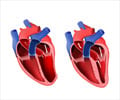
Hypertrophic Cardiomyopathy is usually a familial myocardial disease. About 50 to 60% of cases are due to genetic abnormalities (sarcomeric contractile protein genes to be more specific). It is defined by the presence of hypertrophy (abnormal enlargement) of the left ventricle. Myocardium is the middle muscular layer of heart. A disorderly arrangement of myocytes (muscle cells) occurs in hypertrophic Cardiomyopathy. A number of genetic conditions are associated with this condition.
Hypertrophic Cardiomyopathy presents typically during adolescence. Childhood and the early 20s also serve as the times when the entity presents for the first time.
Hypertrophic Cardiomyopathy is the most common familial genetic disease of the heart with an incidence of - 1/500 to 1/1000.
Mortality rate in adults is about 1%.
Symptoms & Signs
Symptoms may develop at any age. Approximately 30% of adults develop chest pain on exertion.
- Chest pain following a meal (postprandial angina) associated with mild exertion is typical.
- Difficulty with breathing (dyspnoea) is common in adults.
- Paroxysmal nocturnal dyspnoea - here the patient awakens from sleep with acute, severe shortness of breath after 1 to 3 hours of sleep.
- Syncope, i.e. a spontaneous loss of consciousness can occur in 20% of patients.
- Palpitation – this is quite common.
- Sudden death can also be the initial presentation.
Diagnosis
Diagnostic evaluation begins with elicitation of a proper medical history. A detailed family history is taken. This is followed by a careful physical examination. 90% of patients have abnormal ECG findings. The keystone of diagnostic imaging is two-dimensional echocardiography. In case the echocardiogram is of poor quality then the alternatives magnetic resonance imaging (MRI) and computed tomography (CT) are ordered for.
Treatment
Treatment aims at bringing improvement in symptoms and prevention of disease-related complications. Medical modalities include the use of drugs called –
- Beta blockers (â blockers like propranolol),
- Calcium antagonists (verapamil, diltiazem),
- Diuretics (furosemide)
- And Antiarrhythmic drugs like disopyramide.
In patients with significant obstruction in outflow of blood surgical intervention helps. Ventricular septal myectomy is the most commonly done procedure.
Family Screening
12-lead ECG and two-dimensional echocardiography studies are recommended annually during puberty and adolescence and then every 5 years as adults in first degree relatives.
 MEDINDIA
 Email
Email





Which age group people are more affected by Cardiomyopathy?