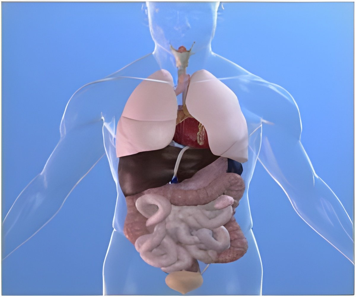
The idea of building a three-dimensional artificial capillary system is that, it can keep living cells viable and functional for more than a week.

TOP INSIGHT
The artificial capillary system made using the cotton-candy spinning method can keep living cells viable and functional for more than a week.
"Some people in the field think this approach is a little crazy," said Bellan, "But now we’ve shown we can use this simple technique to make microfluidic networks that mimic the three-dimensional capillary system in the human body in a cell-friendly fashion. Generally, it’s not that difficult to make two-dimensional networks, but adding the third dimension is much harder; with this approach, we can make our system as three-dimensional as we like."
Many tissue engineering researchers, including Bellan, are currently focusing their efforts on a class of materials similar to hair gel - water-based gels, called hydrogels - and using these materials as scaffolds to support cells within three-dimensional artificial organs.
Hydrogels are attractive because their properties can be tuned to closely mimic those of the natural extracellular matrix that surrounds cells in the body. Unlike solid polymer scaffolds, hydrogels support diffusion of necessary soluble compounds; however, oxygen, nutrients and wastes can only diffuse a limited distance through the gel. As a result, cells must be very close (within the width of human hair) to a source of nutrients and oxygen and a sink for the wastes they produce, otherwise they starve or suffocate.
So, to engineer tissues that have the thickness of real organs and keep cells alive throughout the entire scaffold, the researchers must build in a network of channels that allow fluids to flow through the system, mimicking the natural capillary system.
In the bottom-up process, scientists culture cells in a thin slab of gel, and after some time they spontaneously begin creating capillaries. Although this approach has the advantage of simplicity, it has one fundamental problem: It can take weeks for the cells to create such a network. So it isn’t possible to stack the cells too high or the ones in the center begin dying off before the crucial capillary network forms.
As a result, Bellan is using a top-down approach. He reports that his cotton-candy spinning method can produce channels ranging from three to 55 microns, with a mean diameter of 35 microns. "So far the other top-down approaches have only managed to create networks with microchannels larger than 100 microns, about ten times the size of capillaries," he said. In addition, many of these other techniques are not able to form networks as complex as the cotton candy approach.
Bellan’s focus on this unique use of a cotton candy machine dates back to graduate school. At the time, he was doing research on electrospinning, a process of making nanofibers using strong electric fields. He went to a lecture on tissue engineering where the speaker discussed the need to create an artificial vascular system to support cells in thick engineered tissue. He realized that electrospinning can make networks somewhat resembling capillaries, but at a much smaller scale.
However, getting from that point to creating artificial capillaries that work was not a simple matter. If you create a network of fibers using sugar, when you pour a hydrogel on it, the sugar dissolves away because the hydrogel is mostly water.
This illustrates what Bellan describes as the "Catch-22" in creating such sacrificial structures. "First, the material has to be insoluble in water when you make the mold so it doesn’t dissolve when you pour the gel. Then it must dissolve in water to create the microchannels because cells will only grow in aqueous environments," he explained.
The researchers experimented with a number of different materials before they discovered one that worked. The key material is PNIPAM, Poly(N-isopropylacrylamide), a polymer with the unusual property of being insoluble at temperatures above 32 degrees Celsius and soluble below that temperature. In addition, the material has been used in other medical applications and has proven to be rather cell-friendly.
The researchers first spin out a network of PNIPAM threads using a machine closely resembling a cotton candy machine. Then they mix up a solution of gelatin in water (a liquid at 37 degrees) and add human cells, like adding grapes to jello. Adding an enzyme commonly used in the food industry (transglutaminase, nicknamed "meat glue") causes the gelatin to irreversibly gel. This warm mixture is poured over the PNIPAM structure and allowed to gel in an incubator at 37 degrees. Finally, the gel containing cells and fibers is removed from the incubator and allowed to cool to room temperature, at which point the embedded fibers dissolve, leaving behind an intricate network of microscale channels. The researchers then attach pumps to the network and begin perfusing them with cell culture media containing necessary chemicals and oxygen.
"Our experiments show that, after seven days, 90 percent of the cells in a scaffold with perfused microchannels remained alive and functional compared to only 60 to 70 percent in scaffolds that were not perfused or did not have microchannels," Bellan reported.
Now that Bellan and his team have shown that this technique works, they will be fine-tuning it to match the characteristics of the small vessel networks in different types of tissues, and exploring a variety of cell types.
"Our goal is to create a basic ‘toolbox’’ that will allow other researchers to use this simple, low-cost approach to create the artificial vasculature needed to sustain artificial livers, kidneys, bone and other organs," Bellan said.
Co-authors on the paper are postdoctoral researchers Jung Bok Lee, Xintong Wang, and Shannon Faley, doctoral students Bradly Baer and Daniel Balikov, and Associate Professor of Biomedical Engineering Hak-Joon Sung.
Source-Medindia
 MEDINDIA


 Email
Email


