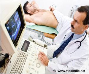
Ultrasound-assisted optical imaging technology has been developed, which could replace endoscopy. The technology is non-invasive, painless and patient-friendly, which could revolutionize medical imaging.
- Ultrasound-assisted optical imaging technology has been discovered
- The technology is completely non-invasive, easy to use, and patient-friendly
- It could replace existing invasive procedures such as endoscopic imaging
The study was led by Dr. Maysam Chamanzar, Ph.D, who is an Assistant Professor of Electrical and Computer Engineering at Carnegie Mellon University, Pittsburgh, Pennsylvania, USA. The study was conducted by Matteo Giuseppe Scopelliti, who is a Ph.D candidate at Carnegie Mellon University’s College of Engineering.
The study has been published in the journal Light: Science & Applications, which is a Springer Nature publication.
TOP INSIGHT
Ultrasound-assisted optical imaging technology has been developed, which could replace invasive procedures like endoscopy. The technology is non-invasive, painless and patient-friendly and could revolutionize the medical imaging field.
What are the Disadvantages of Endoscopic Imaging?
The main disadvantage of endoscopic imaging is that it is highly invasive. In this procedure, an endoscope consisting of a thin tube or catheter, fitted with a miniature camera and biopsy instruments need to be inserted deep within the body cavities for establishing a diagnosis. This procedure is very awkward and uncomfortable, making it unpopular among patients.What is Novel about the New Imaging Technology?
The novelty of the new imaging technology lies in the fact that it permits visualization of deep tissues within the body, which cannot be visualized by light. Since the technique is non-invasive, it is essentially a painless procedure, which makes it patient-friendly as well as easy to use. Moreover, by using ultrasound, a ‘virtual lens’ is created within the body, which alleviates the need for implantation of a ‘physical lens,’ thereby making the procedure much less cumbersome and more user-friendly. This novel technology has allowed the visualization of deeply situated tissues, which has never before been possible using non-invasive techniques.What is the Underlying Mechanism Involved?
It is an established fact that biological tissues block light waves, especially those falling within the visual range of the spectrum. Consequently, currently available imaging techniques cannot use visible light to visualize deep-seated tissues within the body.The new technology, on the other hand, uses ultrasound waves, which are capable of compression and rarefaction of the medium through which they pass. This alters the speed of light, which travels slower in compressed regions and faster in the rarefied regions. This results in the creation of a ‘virtual lens’ that focuses light within the tissue, allowing greater transparency.
The focusing accuracy of this ‘virtual lens’ can be fine-tuned by altering the parameters of the ultrasound waves, allowing visualization of tissues located at different depths within the body. Moreover, the ‘virtual lens’ can be moved about without disturbing the medium by controlling the properties of the ultrasound waves from outside, allowing visualization non-invasively.
“Being able to relay images from organs such as the brain without the need to insert physical optical components will provide an important alternative to implanting invasive endoscopes in the body,” says Chamanzar. “We used ultrasound waves to sculpt a virtual optical relay lens within a given target medium, which, for example, can be biological tissue. Therefore, the tissue is turned into a lens that helps us capture and relay the images of deeper structures. This method can revolutionize the field of biomedical imaging.”
What are the Applications of the New Technology?
Some of the potential applications of this new technology are listed below:- Brain tissue imaging for monitoring brain activity
- Dermal imaging for diagnosing skin diseases
- Cancer imaging for detecting malignant tumors
- Optical imaging in computer vision
- Metrology (the science of measurement)
- Industrial imaging applications at the micron scale
What are the Future Prospects?
Ultrasound-assisted optical imaging technique appears to have a bright future ahead. The technology can be used as a handheld device or a wearable skin patch, depending on which organ is being imaged. This will enable the physician to instantly visualize what’s going on inside the body, without having to do an endoscopy. Indeed, endoscopy may even be replaced by this new technology in the future. Moreover, this new acousto-optic technology could be used for research purposes involving brain imaging to better understand conditions such as Parkinson’s disease, as well as psychiatric and other brain disorders.Concluding Remarks
“What distinguishes our work from conventional acousto-optic methods is that we are using the target medium itself, which can be biological tissue, to affect light as it propagates through the medium,” says Chamanzar. “This in situ interaction provides opportunities to counterbalance the non-idealities that disturb the trajectory of light.”Scopelliti concludes: “Turbid media have always been considered obstacles for optical imaging.” He adds: “But we have shown that such media can be converted to allies to help light reach the desired target. When we activate ultrasound with the proper pattern, the turbid medium becomes immediately transparent. It is exciting to think about the potential impact of this method on a wide range of fields, from biomedical applications to computer vision.”
The research team is optimistic that the new imaging technology could be available for clinical use within a matter of five years.
Reference:
- Breakthrough in optical endoscopy using ultrasound - (https://engineering.cmu.edu/news-events/news/2019/07/16-chamanzar-nature.html)
Source-Medindia
 MEDINDIA


 Email
Email


