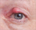
An objective eye test can detect long COVID because it is linked to symptoms of pain, light sensitivity, irritation, and a vague pressure sensation in the eyes.
- Long COVID affects around 1 in 10 people who recover from COVID-19
- A link between changes in cornea, the transparent outer layer of the eye and long COVID identified
- The dysfunction or damage of one or more nerves in long COVID may be the underlying cause of corneal nerve damage
- Evaluating these eye symptoms using corneal confocal microscopy can help predict the severity of long COVID
TOP INSIGHT
People suffering from long COVID have corneal nerve fiber damage and increase in immune cells in the eyes.
Read More..
Long COVID
Encountering with the SARS-CoV-2 coronavirus has left several survivors with long-term symptoms called 'long COVID'. It is estimated that 1 in 10 develops long COVID, where symptoms continue for weeks or months after the acute phase of COVID-19 has passed.The symptoms of long COVID include tight chest, coughing, tiredness, palpitations, feeling breathless, muscle pain, or difficulty focusing.
Researchers have also noted neurological symptoms, such as headache, loss of taste or smell, numbness, dizziness, and pain due to problems with the nervous system in long COVID.
Now, scientists suggest that loss of nerve fibers and the rise in the number of key immune cells in the eye may also serve as an indicator of long COVID, especially in people with neurological symptoms.
Eye Nerve Damage
Researchers think that COVID-19 can transmit SARS-CoV-2 coronavirus directly to a person’s nervous system.They also suggested that neuropathy, the dysfunction or damage of one or more nerves, may be the underlying cause of long COVID induced small nerve damage in the cornea of the eyes. Cornea is the eye's clear and protective outer layer at the front part.
Corneal nerve damage is associated with symptoms of pain, light sensitivity, irritation and sometimes a vague sensation of pressure. Crucial daily activities like reading, driving, and computer work can be affected.
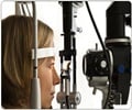
To analyze the damage in cornea scientists used a non-invasive and high-resolution imaging technique known as corneal confocal microscopy (CCM).
CCM is a rapid way of identifying eye nerve damage and inflammation associated with wide range of peripheral and central neurodegenerative conditions like multiple sclerosis, diabetic neuropathy and fibromyalgia.
The scientists also observed the increase in density of dendritic cells, which are immune cells responsible for triggering adaptive immune responses apart from revealing the nerve damage.
Neurological Symptoms Link
In a recent study, 40 people who had COVID-19 during the past 1–6 months and 30 people to act as a control were recruited.The 40 people who had recovered from COVID-19 were asked to complete a questionnaire to identify neurological symptoms at 4 and 12 weeks after receiving a diagnosis of the disease.
It consisted of 28 questions and it covered generalized, cardiovascular, dermatological, gastrointestinal, musculoskeletal, neurological, psychological/psychiatric, respiratory, and ear, nose and throat symptoms. The total score ranged from 0 to 28.
They also found that neurological symptoms were present in 22 out of 40 participants (55 per cent) at four weeks, and 13 out of 29 patients (45 per cent) at 12 weeks.
Later, all of the participants underwent corneal confocal microscopy and they compared the scans of the control group and the participants who had recovered from COVID-19.
Researchers found that the level of corneal nerve fiber damage and number of dendritic cells was greater in individuals having neurological symptoms at 4 weeks. This suggests a strong correlation between the long COVID and corneal nerve fiber damage.
Among those who did not have neurological symptoms, the number of corneal nerve fibers was similar to control group. However, the number of dendritic cells was higher in their case.
Limitations
The study is bound by certain limitations. As it was an observational study, the cause was not established. The size of the sample was relatively small, long-term monitoring was absent, and there was also a dependence on questionnaires to confirm how severe the neurological symptoms were instead of more objective methods.Dr. Malik said, “Larger studies are required to confirm our initial findings, which show definite corneal nerve damage in patients with long COVIDand an ongoing immune response long after these patients have ‘recovered’ from acute COVID-19. We also need to perform longitudinal studies to see the evolution of this condition.”
Despite its limitations, the research demonstrates the fact that eye health is closely linked to overall health and corneal confocal microscopy may have clinical utility as a rapid objective ophthalmic test to evaluate long COVID patients in the future.
Treatment
Damaged nerves usually regenerate on their own but untreated inflammation can make damaged nerves more sensitive.Consult an ophthalmologist immediately to address the injury and inflammation. If not, over time they can trigger inappropriate signals transmitted to and from the brain, resulting in more severe generalized pain.
Treatments can include:
- Eye drops made with the patient’s own blood (autologous serum tears)
- Low-dose anti-inflammatory steroids
- Amniotic membrane lenses
- Neurostimulation
- Blue filter glasses
- Systemic neuro-modulatory therapy
- Topical recombinant corneal nerve growth factors
Reference:
- What Is Neuropathic Corneal Pain? - (https://www.aao.org/eye-health/diseases/what-is-neuropathic-corneal-pain-2)
Source-Medindia
 MEDINDIA



 Email
Email

