- American Academy of Ophthalmology - Preferred Practice Patterns – Age Related Macular Degeneration
- All India Ophthalmological Society CME series - Age Related Macular Degeneration
- Cammaroto, Simona, et al. "Charles Bonnet syndrome." Functional neurology23.3 (2008): 123
- Wong, Wan Ling, et al. "Global prevalence of age-related macular degeneration and disease burden projection for 2020 and 2040: a systematic review and meta-analysis." The Lancet Global Health 2.2 (2014): e106-e116.
- Lu X, Sun X. Profile of conbercept in the treatment of neovascular age-related macular degeneration. Drug Design, Development and Therapy. 2015;9:2311-2320. doi:10.2147/DDDT.S67536.
- Treat-and-Extend Strategy: Is There a Consensus? - (https://www.aao.org/eyenet/article/treat-extend-strategy-is-there-consensus)
What is Age Related Macular Degeneration?
The retina is the light sensitive neural membrane at the back of the eye. It consists of 10 layers, of which the outermost is the retinal pigment epithelium (RPE). External to the RPE is the Bruch’s membrane, which lines the choroid (vascular membrane that forms the posterior part of the uveal tract, and which lies between the sclera and retina).
The macula is the central portion of the retina and measures about 5 mm in size. In the center of the macula is the fovea (1.9 mm in diameter), in the center of which is a depression containing the foveola. The fovea is only about half as thick as the rest of the retina (0.37 mm) and the foveola is 0.13 mm thick. The cones (which are responsible for sharpness of vision and color vision.) in the fovea are larger in size and greatest in number compared to any other part of the retina.
The macula is thus that part of the retina that is required for the greatest acuity of vision.
Age related age related macular degeneration or age related maculopathy is, as the name suggests, a degeneration of the central part of the retina referred to as the macula, which usually occurs over the age of 50 years.
It is characterized by one or more of the following findings on retinal examination (fundoscopy)
| No. | Finding | Description |
| 1. | Drusen | Yellow deposits between RPE and underlying choroid, which may be small, intermediate or large |
| 2. | RPE abnormalities | Increased or decreased pigmentation |
| 3. | Reticular pseudo drusen | Deposits resembling drusen, underneath the retina |
| 4. | Geographic atrophy of RPE | A sharply demarcated round or oval area of degeneration of the RPE |
| 5. | Neovascular maculopathy(exudative maculopathy) |
|
Neovascular maculopathy is often referred to as wet or exudative maculopathy. The presence of other findings is referred to as dry maculopathy.
How is Age Related Macular Degeneration (AMD) Classified?
The age related eye disease study (AREDS) has classified AMD into early, intermediate and advanced AMD. Characteristics of each of these stages are shown in the table below:
| Stage | Characteristics |
| Early AMD | Small drusen, few intermediate drusen or RPE abnormalities |
| Intermediate AMD | Numerous intermediate drusen, geographic atrophy not involving center of fovea |
| Advanced AMD | Geographic atrophy of foveal centerNeovascular maculopathy |
Geographic atrophy is the most advanced form of dry maculopathy, and neovascularization constitutes wet maculopathy. Both of these cause severe visual loss. Drusen > 63 microns in size and pigmentary changes are associated with the greatest risk for progression to advanced AMD over a period of 5 years.
Facts and Statistics on Age Related Macular Degeneration (AMD)
- AMD accounts for about 8.7 % of the world’s blindness and is more common in developed countries, where it is among the leading causes of irreversible blindness. The number of people affected may increase as the percentage of aging population increases.
- 80% of patients diagnosed with AMD have the non-neovascular type; however, neovascular (or wet AMD or exudative AMD) accounts for 90% of severe visual loss.
What are the Risk Factors for Age Related Macular Degeneration (AMD)?
The exact cause of AMD remains unclear but the following risk factors predispose to the condition.
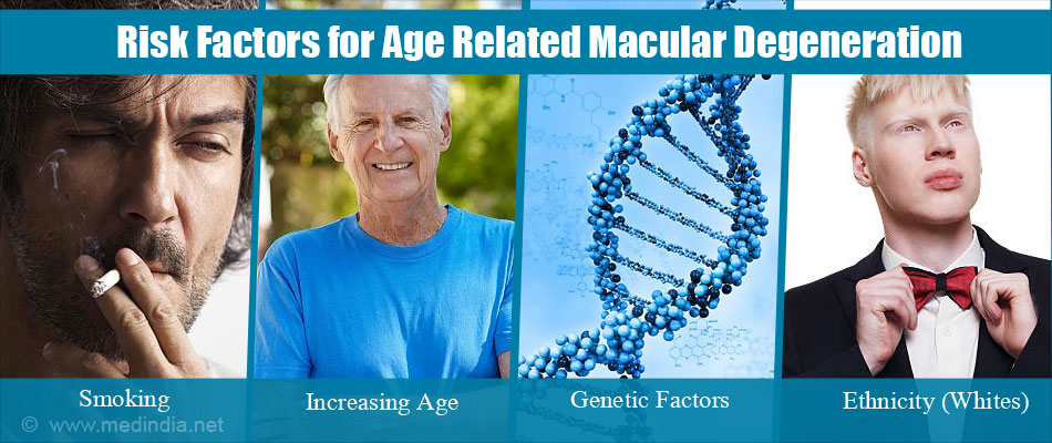
- Increasing age: AMD usually occurs in persons over the age of 50 years, although some of the findings that are precursors of this condition are seen between the ages of 40 and 50 years. The risk increases with each decade after 50 years, with the highest prevalence in the > 80 year age group. Most of the population bases studies have noted a threefold increase in prevalence in persons aged > 75 years compared to those < 50 years old.
- Ethnicity: Prevalence of both non-exudative and exudative AMD is more common in whites as compared to other races.
- Genetic factors. A family history of AMD is a significant risk factor.
- Cigarette smoking: Smoking has been found to be the main modifiable risk factor for AMD; numerous studies have consistently identified the same. Cessation of smoking lowers the risk, and those who have stopped smoking for 20 years have the same risk as non-smokers. Lifetime exposure to cigarette smoke (which takes into account number of packs of cigarettes per day, and duration of smoking) is significant for assessing risk.
- The Beaver Dam Eye Study (which studied the relationship between smoking and the natural history of AMD) showed that –
- Current smoking and a greater lifetime exposure to smoking caused greater progression of AMD
- A greater lifetime exposure to smoking was associated with a greater incidence of early and late AMD
- Current smoking and a greater lifetime exposure to smoking were associated with an increased risk of death
- Low systemic levels of antioxidants such as vitamins C, E, carotenoids (lutein and zeaxanthin) and zinc
- Obesity or a high body mass index
- An increased risk of AMD was found in persons with a diet high in saturated fats and cholesterol, and a reduced risk with a higher intake of food rich in omega-3 fatty acids such as fish.
- A family history of AMD – A strong association with AMD is found on chromosome 10. Other genetic markers predict risk of progression of AMD
| Most Important Risk Factors for Advanced AMD |
|
How Does Age Related Macular Degeneration (AMD) Occur?
The following mechanisms may be involved in its development
- Accumulation of metabolic debris over time leads to retinal pigment epithelial (RPE) dysfunction. This RPE dysfunction leads to drusen formation and in later stages to the growth of abnormal choroidal blood vessels (choroidal neovascularization).
- Release of Vascular endothelial growth factor (VEGF) in response to tissues getting less oxygen; it promotes the growth of new and abnormal blood vessels. These new vessels grow through breaks in Bruch’s membrane and result in bleeding and protein leak underneath the macula.
What are the Symptoms of Age Related Macular Degeneration (AMD)?
The symptoms depend on the specific findings that are present.
- Gradual progressive diminution of vision. This is a feature of the dry type of maculopathy.
- Rapid loss of vision – this is characteristic of wet maculopathy
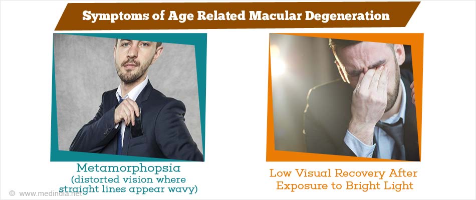
- Metamorphopsia – distorted vision where straight lines appear wavy
- Central scotoma – shadows seen in the central part of vision
- Difficulty in hue discrimination
- Slow visual recovery after exposure to bright light
- Diminished contrast sensitivity
- New symptoms suggestive of neovascularization are the sudden appearance of shadows, waviness of lines and diminished vision.
How do you Diagnose Age Related Macular Degeneration (AMD)?
AMD is diagnosed by a thorough history as well as eye examination given below:
How is a Patient with Age Related Macular Degeneration (AMD) Examined and Evaluated?
A thorough ophthalmic examination is carried out including checking visual acuity, refraction and slit lamp examination.
- The retina is examined using slit lamp biomicroscopy and binocular indirect ophthalmoscopy for characteristic dry and wet AMD changes
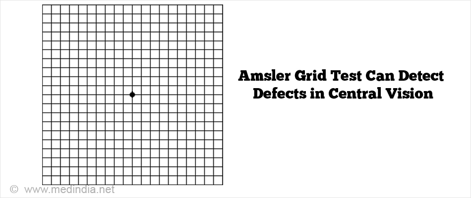
- Amsler grid test – This test checks for defects in central vision. The Amsler grid consists of a pattern of intersecting straight lines with a black dot in the center for maintaining fixation. The person should cover one eye, and, with the other eye fixing on the black dot, note the appearance of the other lines. If the lines or parts of them appear distorted or missing, it indicates a disease of the macula.
- Optical Coherence Tomography (OCT) – an imaging technique that provides high quality cross sectional images of the eye, is now the imaging modality of choice for evaluation of macular lesions. It not only helps in diagnosis, but also helps in evaluation of response to therapy in neovascular AMD
- Fundus fluorescein angiography – this involves injection of an intravenous contrast dye and observing the retinal vasculature through a special fundus camera. It is primarily used in AMD to confirm the presence of neovascularization.
- Indocyanine green angiography – use of indocyaninegreen instead of fluorescein helps in better delineation of choroidal neovascularization, especially in atypical cases.
Fundus fluorescein angiography and indocyanine green angiography are used only in select cases.
How is Age Related Macular Degeneration (AMD) Treated?
The treatment of AMD depends on the stage. The following table briefly explains the guidelines for treatment of various stages of AMD.
The AREDS2* is the Age Related Eye Disease Study2, the results of which were published in 2011 and found that addition of specific antioxidant supplements proved beneficial in intermediate AMD and unilateral advanced AMD (see below)
| Stage | Treatment | Comments |
| Early AMD | No treatment | Annual checkups with the eye doctor are recommended to detect progression. Only 1.3 % of people in this stage progress to advanced AMD in 5 years. However, immediate consultation if there are new symptoms suggestive of neovascularization |
| Intermediate AMD | AREDS2 formula of vitamin and mineral supplementation | Daily supplementation with this combination reduced progression to advanced AMD at 5 years by 25%, and reduced the risk of losing 3 or more lines of vision by 19%. Immediate consultation if there are new symptoms suggestive of neovascularization |
| Advanced non-neovascular AMD | Observation if bilateral,AREDS2 supplements if unilateral | Annual checkups with the eye doctor are recommended to detect progression. However, immediate consultation if there are new symptoms suggestive of neovascularization |
| Neovascular AMD | Intravitreal injection of anti-VEGF agents | Injection into the vitreous of anti-VEGF agents like ranibizumab, bevacizumab and aflibercept are the first line treatment for neovascular AMD today |
| Photodynamic therapy with verteporfin | In cases of new vessels underlying the fovea, that do not respond to intravitreal anti- VEGF agents. This involves intravenous injection of a photosensitizing drug (verteporfin) and then treating the eye with a laser, the verteporfin in the blood vessels of the eye absorbs the laser, and the blood vessels are destroyed. | |
| If unilateral, treatment with AREDS2 supplements reduces progression to advanced AMD in the fellow eye |
| AREDS2* formula of vitamin and mineral supplementation | |
| Supplement | Daily dose |
| Vitamin C | 500 mg |
| Vitamin E | 400 IU |
| Lutein | 10 mg |
| Zeaxanthin | 2 mg |
| Zinc oxide | 25 mg |
| Copper oxide | 2 mg |
The frequency of intravitreal injections depend on the treating doctor and there are various regimens available.
| Treatment Regimens for Intravitreal Injections | |
| Fixed continuous treatment regimen | Fixed schedule of injections every 4-8 weeks |
| PRN (Pro Re Nata or as needed) | After the initial injection, monthly clinical exams and OCT are done, but whether or not to inject is decided depending upon the response and OCT findings |
| Treat and extend (popular today as the number of visits is less than the other 2 regimens) | After a certain number of monthly injections, the intervals between visits is extended, usually by 2 weeks at a time up to a maximum of 12 weeks intervals. Injections are given at every visit. |
A new drug, conbercept has been approved in China. It has been found to be effective when used as quarterly injections (every 3 months).
How you can Cope with Advanced Central Visual Loss
You will have to make use of the surrounding area of the retina which has relatively good vision to help in your tasks. This may be challenging at first, but with consistent practice, it can be achieved. Also improving the ambient lighting and use of magnification go a long way in easing day to day near tasks. In addition, discuss with your doctor if you are feeling depressed. Depression can make the effects of AMD worse.
Some recommendations are:
- Carry a small lamp or flashlight with you at all times
- Increase contrast by using felt tip or gel pens instead of ball point pens for writing. Use a white cup for coffee or tea.
- Sit closer to the television for better view
- Get books, keyboards, cell phones and TV remotes with large print.
- Use large sized font on your computer. Use an e-reader or electronic tablet for reading.
- For printed material, use a lighted hand held magnifier, stand magnifier or video magnifier.
- Organize your daily requirement with specific places designed for specific items for ease of access
- Make use of tactile stimuli in the form of safety pins, rubber bands and clips.
- Use audio formats where possible for telling time, calculator and books.
- Join a social group with similar issues
How do you Prevent Age related Macular Degeneration?
Patients with early AMD or a family history of AMD should go in for regular eye exams to detect the intermediate stage, which, if present, can be treated with supplements to reduce the risk or progression to advanced AMD.
Regular home testing with an Amsler grid (in each eye individually) can help detect early onset of neovascular maculopathy and thus early consultation and treatment can prevent more severe forms of visual loss. Any form of visual deterioration, shadow formation, waviness of lines or missing lines should be immediately reported to your doctor.
It is strongly recommended for patients to give up smoking.
Though there is no concrete evidence for the same, ultraviolet light is believed by some to be a risk factor. Wear sunglasses when you go outdoors.

Health tips:
- Eat plenty of green leafy vegetables and fish
- Reduce the intake of cholesterol and saturated fats
- Maintain healthy weight for your height
- Wear sunglasses while outdoors
- Quit smoking
- Go for regular eye examinations after the age of 40
 MEDINDIA

 Email
Email


