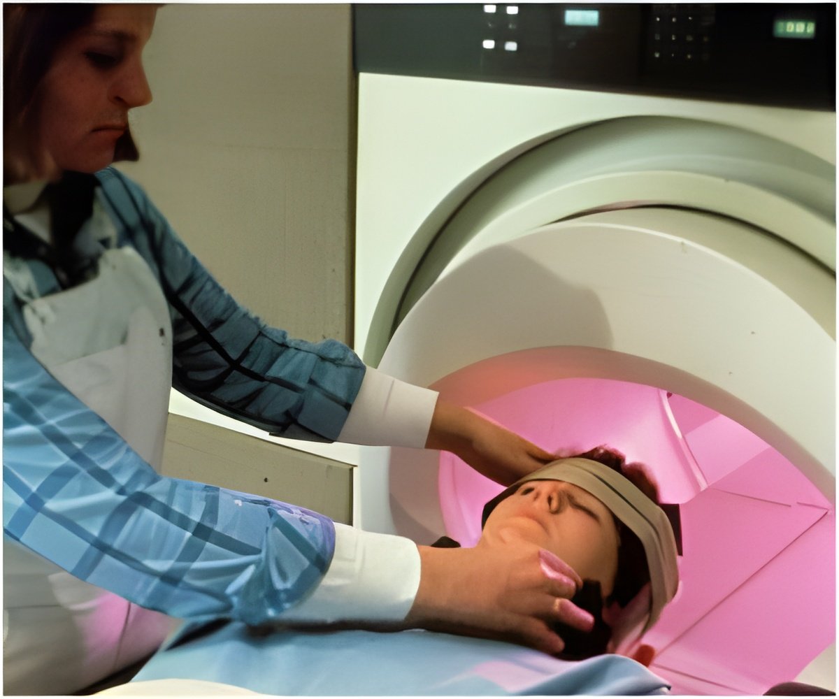
Brain imaging after mild traumatic brain injury (mTBI) or a concussion can detect tiny lesions, which may prove to be a target for treating people with mTBI.

The study involved 256 people with an average age of 50 who were admitted to the emergency department at Suburban Hospital in Bethesda and Washington Hospital Center in the District of Columbia after mild head injuries. They underwent magnetic resonance imaging (MRI) brain scans. Of those, 104 had imaging evidence of hemorrhage in the brain (67 percent reported loss of consciousness, and 65 percent reported amnesia, or temporary forgetfulness). People with hemorrhages underwent more detailed brain scans with advanced MRI within an average of 17 hours after the injury.
Advanced imaging showed that—of those 104 people with imaging evidence of hemorrhage—20 percent had microbleed lesions and 33 percent had tube-shaped linear lesions. Microbleeds were distributed throughout the brain whereas linear lesions, which were found mainly in one area, were more likely to be associated with injury to adjacent brain tissue.
The investigators hypothesized that the linear lesions seen on MRI may represent a type of vascular injury that is seen in brain tissue studies of people with more severe TBI. "If that theory is confirmed, it may provide an opportunity to develop treatment strategies for people who have suffered a mild TBI," said Parikh.
 MEDINDIA


 Email
Email


