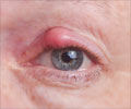
Examination of the eyes with a special dye could give clues on the extent of brain damage caused due to stroke in the future.
- Gadolinium contrast used during MRI in stroke patients leaks into the eyes.
- This indicates that the blood vessels of the eyes become leaky following a stroke.
- Therefore, an eye examination following a stroke using a special dye could obviate the need for an MRI scan in the future.
- Leakage of gadolinium occurred into the eyes of 76% individuals.
- There also appeared to be a specific pattern for the leakage. The MRI done after 2 hours showed leakage in the aqueous chamber, the part of the eye in front of the iris in 67% individuals, in the vitreous chamber, the part of the eye behind the iris in 6% patients, and in both the aqueous and the vitreous chamber in 27% of patients. Patients with gadolinium in both the chambers had a larger part of the brain affected by the stroke.
- The MRI performed at 24 hours detected the leakage in the vitreous chamber of the eyes in 75% patients, and in both the chambers in 6% patients. In older patients, patients with greater affliction of the white matter of the brain due to aging, and patients with hypertension, gadolinium was detected more commonly in the vitreous chamber at 24 hours.
TOP INSIGHT
Examination of the eyes with a special dye could give clues on the extent of brain damage caused due to stroke in the future.
Further studies on the phenomenon could reveal ways in which, using a special dye that accumulates in the eye in stroke patients similar to gadolinium, an eye examination will be able to provide clues into the severity of brain damage caused by the stroke.
About MRI
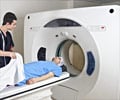
Magnetic resonance imaging (MRI) is an imaging test during which images of the brain or other parts of the body are obtained using a magnetic field. A contrast is sometimes injected to highlight the damaged part. In a normal brain, leakage of the dye through the blood vessels into the brain does not occur. Damage to the brain on the other hand, allows the contrast to enter the brain and settle down in damaged areas, thus enabling easy detection of the damaged parts.References:
- Hitomi et al. Blood-ocular barrier disruption in acute stroke patients. Neurology. February 7, 2018. doi: 10.1212/WNL.0000000000005123.
 MEDINDIA


 Email
Email


