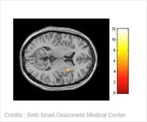
Measuring markers of bone injury in the blood can help predict the likelihood of a cranial lesion and determine the need for head computed tomography, in cases of traumatic brain injury.

The authors, Linda Papa and colleagues from Orlando Regional Medical Center, North Florida Veteran's Health System and University of Florida (Gainesville), University of Central Florida (Orlando), Banyan Biomarkers Inc. (Alachua, FL), Virginia Commonwealth University (Richmond, VA), and Baylor College of Medicine (Houston, TX), showed that increased blood levels of glial fibrillary acidic protein (GFAP) following TBI was a good predictor of intracranial lesions, whether or not the patient had fractures elsewhere in the body. Whereas S100ß levels in the blood of were significantly higher in trauma patients with fractures than without fractures, it was not as useful as GFAP in distinguishing between intracranial and extracranial lesions.
John T. Povlishock, PhD, Editor-in-Chief of Journal of Neurotrauma and Professor, Medical College of Virginia Campus of Virginia Commonwealth University, Richmond, notes that "This is an extremely important paper because of its relatively large sample size and its singular focus upon mild traumatic brain injury complicated by the presence of extracranial lesions. This study convincingly demonstrates the efficacy and brain specific nature of GFAP and its ability to detect traumatic intracranial lesions while also calling into question the overall utility of S100ß in the same patient population. Importantly, the superior performance of GFAP in the mild brain injured population is an important observation consistent with other reports emerging in the field. Lastly, the observation that these GFAP elevations occur relatively early in a post-traumatic course speaks to the potential utility of using these biomarkers to screen brain injured patients who then may require more extensive and/or long term imaging studies."
Source-Eurekalert
 MEDINDIA


 Email
Email

