Diagnosis
Diagnosis of rhabdomyosarcoma is difficult and is often delayed because the symptoms are not very evident.
Early diagnosis is vital to control the disease that spreads quickly.
The following tests may be carried out in a patient with suspected rhabdomyosarcoma
- Tumor Biopsy
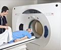
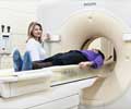
- CT scan /MRI scan of the site of tumor
- CT scan of the chest to check for metastasis
- Bone marrow biopsy /bone scan to check for cancer spread
- Spinal tap (lumbar puncture)
The protein myo D1 is not found in normal skeletal muscle but is found in rhabdomyosarcoma. It functions as an immunohistochemical marker forthis tumor.
When rhabdomyoblasts are viewed under a light microscope, striations are visible. The subcellular changes, however, can only be detected with the help of an electron microscope.
Based on the types of the cells, rhabdomyosarcoma tumors are classified as any one of the following:
Embryonal -This type of tumor is common among children under the age of 15 and usually occurs in the regions of the head, neck or the genitourinary tract. This form of rhabdomyosarcoma is most responsive to treatment than any other type.
Botryoid - This type is a variant of the embryonal variety. Here, the tumor presents itself as a grape-like lesion in hollow organs such as the urinary bladder and the vagina.
Alveolar type- An aggressive tumor, usually involving the muscles of the extremities or trunk.
Pleomorphic or Undifferentiated type- Usually found in adults, this form of tumors arises in muscles of the extremities.


