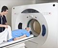
- Ultra-high-strength MRI may release mercury from amalgam dental fillings
- Amalgam fillings contain a high percentage of mercury
- The mercury leak was insignificant in lower strength MRI scanners
MRI scanners use strong magnetic fields and radio waves (radiofrequency energy) to generate images that provide information that may be useful in determining a diagnosis. The 7T MRI has more than double the static magnetic field strength than earlier MRIs where increased strength relates to an equally better overall image quality of MRI. The magnetic field strength in the MRI scanners are measured in Tesla or “T.” Earlier clinical MRI systems were available in field strengths of 3T and below.
Amalgam fillings are otherwise known as silver fillings and have been used in dentistry for many years. Amalgam consists of approximately 50 percent mercury; mercury, as we all know, is a toxin that can cause a host of harmful effects in humans.
The American Dental Association reports that approximately 100 million amalgam filling procedures are performed every year in the U.S. Although the U.S. Food and Drug Administration considers amalgam fillings safe for adults and children older than age six, the use of dental amalgam, however, remains rather controversial due to its high mercury content.
Amalgam fillings are currently banned or restricted in Sweden, Norway, Denmark, Germany, Finland, Netherlands, and Japan.
Connection between MRI and Mercury
"In a completely hardened amalgam, approximately 48 hours after placing on teeth, mercury becomes attached to the chemical structure, and the surface of the filling is covered with an oxide film layer," said the study's lead author, Selmi Yilmaz, Ph.D., a dentist and faculty member at Akdeniz University in Antalya, Turkey. "Therefore, any mercury leakage is minimal."However, the magnets in the stronger scanners cause corrosion in the amalgam, which allows toxic mercury to leak out.
In fact, studies done earlier have revealed that exposure to the magnetic fields of MRI could cause mercury to leak from amalgam fillings, with approximately 40 percent of it traveling into saliva and the gastrointestinal system and around 10 percent of the mercury being absorbed. Also, approximately 60 percent is released as mercury vapor and is either inhaled, entering the lungs, or is exhaled.
The concern increased some more when the ultra-high-strength 7-T scanners were cleared for use by the FDA in the clinic. Yes, the stronger magnetic field of 7-T MRI yields more anatomical detail, but the question remained as to what effect these powerful fields will have on amalgam dental fillings.
"To our knowledge, no previous study has tested the effects of 7-Tesla MRI on mercury release from amalgam fillings," the authors wrote. "We hypothesized that 7-Tesla MRI can trigger mercury release."
Study design - Evaluating mercury released from the dental amalgams after exposure to 7-T and 1.5-T MRIs
Dr. Yilmaz and Mehmet Zahit Adi, Ph.D., studied the teeth of patients that had been extracted for clinical indications.
The researchers opened two-sided cavities in each tooth and applied amalgam fillings.
After nine days, two groups of 20 randomly selected teeth were separated and placed in a solution of artificial saliva
Immediately following this, one group was exposed to 20 minutes of 1.5-T MRI while the other was exposed to 20 minutes of 7-T MRI.
The third group was a control group of teeth that was placed in artificial saliva only
Study results
The researchers then analyzed the artificial saliva solution- The mercury content in the 7-T MRI group was 0.67 ± 0.18 parts per million (ppm) compared to 0.17 ± 0.06 ppm in the 1.5-T MRI group
- The mercury content in the control group was 0.14 ± 0.15 ppm
- This increase of 0.67 ppm in the 7-T group was approximately four times the levels found in the 1.5-T group and the control group
At the same time, the authors reassure that no evidence of harmful effects was found in the 1.5-T group and since this is the scanner widely available and commonly used for patient exams, patients with amalgam fillings should erase their concerns about having an MRI exam.
There are three ongoing projects being planned by, Dr. Yilmaz and his group where they will focus on phase and temperature changes of dental amalgam across different magnetic fields.
Mercury Exposure
Every human is exposed to some level of mercury. However, the type and severity of health effects that occur depend on the type of mercury, the dose, the age or developmental stage of the person exposed, the duration of exposure, and the route of exposure.Mercury toxicity is measured as the amount of mercury in the bloodstream (for short-term exposure) or the amount of mercury excreted in the urine relative to creatine (for long-term mercury exposure).
Elemental mercury is a component of amalgam and is absorbed very differently from the one found in fish, methylmercury.
How much a person is exposed to mercury from amalgam restorations depends on the number and size of restorations, composition, chewing and brushing habits, and many other physiological factors.
Mercury exposure is high during filling placement and removal. Mercury vapors are released when chewing for more than 30 minutes and then subside approximately 90 minutes following chewing cessation. Chewing contributes to a daily mercury exposure for those with amalgam fillings.
Reference:
- Mortazavi G, Haghani M, Rastegarian N, Zarei S, Mortazavi SMJ. “Increased Release of Mercury from Dental Amalgam Fillings due to Maternal Exposure to Electromagnetic Fields as a Possible Mechanism for the High Rates of Autism in the Offspring: Introducing a Hypothesis.” Journal of Biomedical Physics & Engineering. (2016) ;6(1):41-46.
Source-Medindia


