Suffering from shortness of breath, fatigue and extreme cold, Todd Dunlap, 62, arrived at Ronald Reagan UCLA Medical Center's emergency room on Aug. 8.

A new grandfather, Dunlap didn't hesitate to choose the second option and underwent the procedure on Aug. 14. A week later, he was home, full of energy and eager to play on the floor with his 9-month-old grandson.
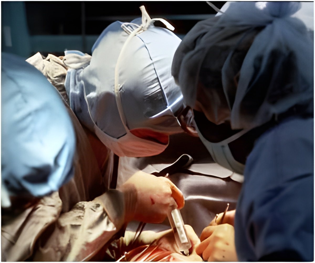
Here's how it worked: A team of UCLA interventional radiologists and cardiovascular surgeons slid a tiny camera down Dunlap's esophagus to visually monitor his heart. Next, they guided a coiled hose through his neck artery and plugged one end into his heart, against the clot. They threaded the other end through a vein at the groin and hooked the hose up to a powerful heart-bypass device in the operating room to create suction.
"Once in place, the AngioVac quickly sucked the deadly clot out of Mr. Dunlap's heart and filtered out the solid tissue," said Moriarty, a UCLA interventional radiologist with expertise in clot removal and cardiovascular imaging. "The system then restored the cleansed blood through a blood vessel near the groin, eliminating the need for a blood transfusion."
The procedure lasted three hours. Doctors observed Dunlap for three days in intensive care before transferring him to the hospital's cardiac ward and then discharging him four days later.
Source-Eurekalert
 MEDINDIA


 Email
Email


