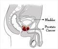
Prostate cancer is the most common type of cancer among men, but its diagnosis has up to now been inaccurate and unpleasant.
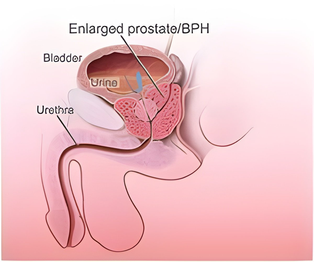
The biopsies also have disadvantages; for example they are not targeted, but instead tissue is sampled randomly using 6 to 12 needles. The chance that the needles will miss a tumor is high, causing a false negative result. In around one-third of cases with negative biopsies, tumors are later found to be present. Furthermore doctors often operate after a positive biopsy, but find a tumor so small that it would have been better not to operate.
The new technology uses the injection of microbubbles of a contrast agent with no side-effects. The response of the tiny bubbles to ultrasound is different from that of human tissue or blood. This makes the bubbles traceable from the outside, right into the smallest blood vessels. The pattern of blood vessels in tumors is different from that in healthy tissue. The researchers can recognize this pattern from advanced analysis of the bubble concentrations. And because tumors need blood – and hence new blood vessels – to grow, the researchers expect to be able to see how aggressive the cancer is from the pattern of the blood vessels.
The technology has been tested on four patients from whom the affected prostate was removed, dr.ir. Massimo Mischi of the TU/e department of Electrical Engineering explains. The location and size of the tumors turned out to match accurately with the images produced using the new technology. Mischi presented these first, promising results at a recent conference in Chicago.
Next year the research team will carry out a pilot with biopsies guided by images made using the new technology. This allows the biopsies to be targeted, and therefore more effective. In a later phase the ultrasound technology will be used to decide whether biopsies are required, which will reduce the number of biopsies carried out. The researchers expect their technology to be available in hospitals within five years. The ultimate goal is for doctors to be able to determine if an operation is necessary, and if so what kind of operation, based on the images produced, without the need for biopsies.
All in all doctors will eventually be able to intervene much more accurately, expects prof.dr.ir. Hessel Wijkstra, head of urology research at AMC Amsterdam. Wijkstra was appointed part-time professor of Hemodynamic Contrast Sonography at TU/e last month. Furthermore he believes that there will be less unnecessary operations. In some cases doctors may decide to leave small, non-aggressive tumors untouched and monitor these tumors, in cases where a tumor is not causing any symptoms and is not impairing the health of the patient. In these cases the health effects of surgery are worse than those of the tumor itself. A further positive effect of this new approach is that the total costs will be reduced.
 MEDINDIA

 Email
Email
![Prostate Specific Antigen [PSA] Prostate Specific Antigen [PSA]](https://www.medindia.net/images/common/patientinfo/120_100/prostate-specific-antigen.jpg)

