A research team from Massachusetts General Hospital developed a collagen-targeting PET Probe which may improve diagnosis and treatment of pulmonary fibrosis.

‘The newly developed collagen-targeting PET probe has been found to improve diagnosis and treatment for pulmonary fibrosis.’


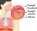
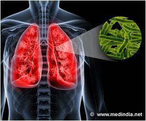
Pulmonary fibrosis is a lung disease which occurs when the lung tissue becomes damaged and scarred. Scientists report how the collagen-targeting probe called 68Ga-CBP8, were bound to scar the tissue in the lungs of two animal models that indicated the extent of fibrosis. It was also able to detect the reduced fibrosis in animals that received the anti-fibrotic drug.Some experiments regarding the human lung tissue also suggested that the probe will be able to differentiate between stable disease and progressive fibrosis.
"Increased collagen production is a hallmark of fibrosis in the lungs and in other organs," explains Peter Caravan, PhD, of the Athinoula A. Martinos Center for Biomedical Imaging and co-director of the Institute for Innovation in Imaging at MGH, a co-corresponding author of the report.
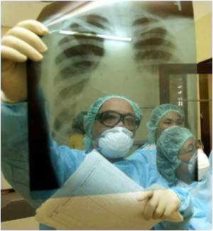
"High resolution CT scanning can precisely diagnose only 50 percent of patients and often cannot predict prognosis or show response to therapies, making invasive biopsy -- which can be hazardous to these patients -- the only definitive diagnostic method. Non-invasive PET imaging with 68Ga-CBP8 provides information on the entire lung without a biopsy, which would only reflect a small portion of the lung."
While pulmonary fibrosis may be caused by exposure to pollutants or result from other disease, in most cases the cause is unknown or idiopathic. The buildup of scar tissue stiffens the lungs, making breathing difficult and restricting the passage of oxygen into the blood. For most patients, the condition progressively worsens, leading to death in 3 to 5 years.
Advertisement
The ability of PET technology to detect and quantify collagen molecules presents significant advantages over CT scanning, which only reveals structural abnormalities, which could be caused by other conditions. The MGH team's initial experiments showed that 68Ga-CBP8 specifically binds to collagen in a mouse model of pulmonary fibrosis but did not accumulate in the lungs of healthy animals.
Advertisement
Application of 68Ga-CBP8 to tissues removed from the lungs of pulmonary fibrosis patients prior to transplantation also showed greater probe accumulation in more heavily scarred areas, suggesting that the results in the mouse models would probably also be seen in human patients.
"The ability of molecular PET imaging with this probe to detect early-stage fibrosis would allow us to begin treatment when it would be most effective," says Michael Lanuti, MD, MGH Division of Thoracic Surgery, co-corresponding author of the Science Translational Medicine paper.
"This probe may also be able to distinguish new, active fibrosis from stable disease, which would allow clinicians to better tailor therapy to individual patients. And since response to therapy is difficult to ascertain with high-resolution CT scanning, PET molecular imaging may be a more sensitive way to detect changes in active fibrosis."
The MGH team is preparing the documentation required to test 68Ga-CBP8 in patients and has received National Heart, Lung and Blood (NHLBI) funding for such a trial, which may begin later this year.
Source-Eurekalert

