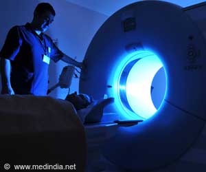
The use of magnetic resonance imaging (MRI) to measure blood flow over atherosclerotic plaques could help identify plaques at a risk of thrombosis, researchers say.

While most studies have focused on the plaque within the vessel wall, the flow of blood in the vessel (hemodynamics) also is known to be important in the progression and disruption of plaques.
In this study the researchers, led by James A. Hamilton, PhD, professor of biophysics and physiology at BUSM, found that the measurement of endothelial sheer stress (ESS), which is the indirect stress from the friction of blood flow over the vascular endothelium surface, can identify plaques in the highest risk category. After performing a non-invasive MRI examination of the aorta in a preclinical model with both stable and unstable plaques, a pharmacological "trigger" was used to induce plaque disruption. Low ESS was associated with plaques that disrupted and had other "high-risk" features, such as positive remodeling, which is an outward expansion of the vessel wall that "hides" the plaque from detection by many conventional methods.
These results are consistent with previous studies that examined coronary arteries of other experimental models using invasive intravascular ultrasound method to measure features of vulnerability but without an endpoint of plaque disruption, which is the outcome of the highest risk plaques.
"Our results indicate that using non-invasive MRI assessments of ESS together with the structural characteristics of the plaque offers a comprehensive way to identify the location of "high-risk" plaque, monitor its progression and assess the effect of interventions," said Hamilton. "Early identification of "high-risk" plaques prior to acute cardiovascular events will provide enhanced decision making and might improve patient management by allowing prompt aggressive interventions that aim to stabilize plaques."
 MEDINDIA


 Email
Email


