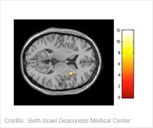
A certain type of scanning can detect when a patient is failing to respond to brain tumour treatment before symptoms appear, according to researchers at Barrow Neurological Institute at St. Joseph's Hospital and Medical Centre.
During the study, researchers analysed patients with recurring malignant brain tumours who were receiving chemotherapy.
Advertisement
They received scans through an imaging device called MR spectroscopy to identify metabolic changes.
The scanning technique suggested that the use of metabolic imaging identifies chemical changes earlier than structural imaging such as a conventional MRI and CT scans.
This approach allowed researchers to determine if the tumours were responding to treatment early by assessing metabolic changes in a brain tumour, which are easy to detect and appear before structural changes or symptoms.
"The study has shown for the first time that metabolic response to brain tumour treatment can be detected earlier and faster by metabolic imaging instead of through structural imaging or assessment of the neurological status of a patient," said Mark C. Preul, M.D., Newsome Chair of Neurosurgery Research at St. Joseph's.
The researchers said that imaging can be done often and poses no radiation hazard. It is also non-invasive.
"Frequent use of this type of imaging may be a useful tool to follow a patient's response to chemotherapy for malignant brain tumours," said Dr. Preul.
"It gives us the ability to identify treatment failure early and more time to alter a patient's treatment plan before the disease progresses," he added.
Source: ANI
RAS/L
Advertisement
Advertisement
|
Advertisement
Recommended Readings
Latest Research News

Relative Energy Deficiency in Sport syndrome is overlooked by athletes and can be worsened by 'sports culture' due to its perceived short-term performance benefits.

New study sheds light on the intrinsic, yet often overlooked, role of sleep preparation as a hardwired survival strategy.

Microgravity-induced bone loss in space, can be reduced by systemic delivery of NELL-1, a protein required for bone growth and its maintenance.

Researchers establish connections between Alzheimer's-linked genetic alterations and the functioning of brain cells.

Team at NeuroRestore introduces a groundbreaking gene therapy that has effectively promoted nerve regrowth and reconnection, post spinal cord injury.