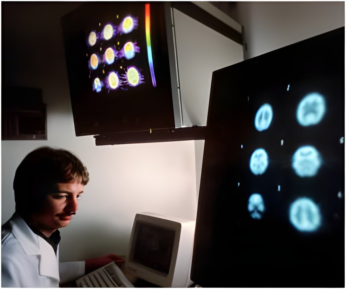
A more complete and continuous two-dimensional original data of nerves can be obtained by high-resolution multilayer X-ray computer tomography.

Jiaming Fu and colleagues from the 98 Hospital of Chinese PLA successfully developed a three-dimensional digital visualization model of healthy human cervical nerves, which overcomes the disadvantages of milling, avoids data loss, and exhibits a realistic appearance and three-dimensional image. Furthermore, vivid images from various angles can be observed due to minimal pattern distortion. This model revealed the morphology, distribution, and spatial relations of the major nerves of the neck, and provided three-dimensional morphological data for anatomical teaching and morphological observation of regenerated nerves, nerve block anesthesia, and surgery. These results are published in the Neural Regeneration Research (Vol. 8, No. 20, 2013).
Source-Eurekalert
 MEDINDIA


 Email
Email


