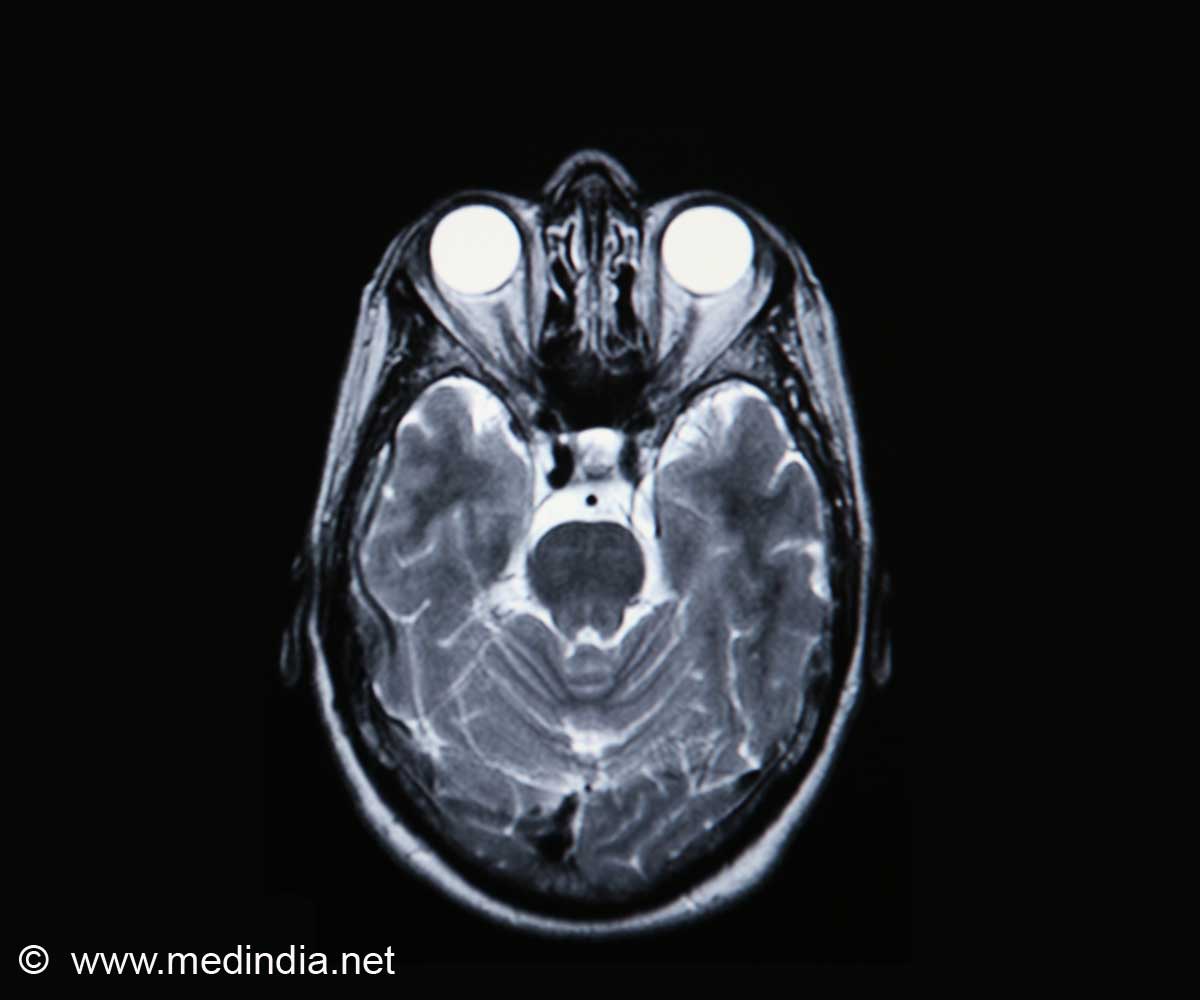
In a noninvasive and non-radioactive manner, magnetic resonance spectroscopy can quantitatively analyze in vivo abnormalities of biochemical metabolism within brain tissue.

In a recent study reported in the Neural Regeneration Research (Vol. 9, No. 4, 2014), 7.0T magnetic resonance spectroscopy showed that in the hippocampus of Alzheimer's disease rats, the N-acetylaspartate wave crest was reduced, and the creatine and choline wave crest was elevated. This finding was further supported by hematoxylin-eosin staining, which showed a loss of hippocampal neurons and more glial cells.
Moreover, electron microscopy showed neuronal shrinkage and mitochondrial rupture, and scanning electron microscopy revealed small size hippocampal synaptic vesicles, incomplete synaptic structure, and reduced number. Overall, these findings from Lei Zhang and co-workers from Beijing Tiantan Hospital Affiliated to Capital Medical University in China revealed that 7.0T high-feld nuclear magnetic resonance spectroscopy detected the lesions and functional changes in hippocampal neurons of Alzheimer's disease rats in vivo, allowing the possibility for assessing the success rate and grading of the animal model of Alzheimer's disease.
Source-Eurekalert
 MEDINDIA


 Email
Email


