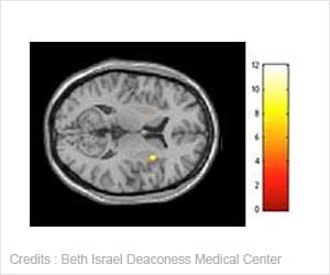
Exhaustion syndrome, also called burnout and exhaustion depression, leaves objectively measurable changes in the brain, a new study from Umea University in Sweden has shown.

The patient group is distinguished by being anxious and pessimistic, with a weak sense of self, which is common in many psychiatric disorders. What was special about this group was that they stood out as persistent, ambitious, and pedantic individuals.
Being ambitious, fastidious, and overachieving also appears to make a person more prone to exhaustion syndrome. According to Agneta Sandstrom's dissertation, individuals with exhaustion syndrome demonstrate impaired memory and attention capacity as well as reduced brain activity in parts of the frontal lobes. Regulation of the stress hormone cortisol is also impacted in the group, with altered sensitivity in the hypothalamic-pituitary-adrenal axis (HPA axis).
The dissertation addresses whether it is possible to use neuropsychological tests to confirm and describe the cognitive problems reported by patients suffering from exhaustion syndrome.
Above all, patients demonstrate problems regarding attention and working memory. Patients were also asked to perform working memory tests while lying in a functional magnetic resonance camera that measures the brains activity patterns.
Exhaustion syndrome patients proved to have a different activity pattern in the brain when they performed a language test of their working memory, and they also activate parts of the frontal lobe less than healthy subjects and a group of patients who had recently developed depression.
There is also a difference in the diurnal rhythm of corisol, with the patients presenting a flatter secretion curve than the other two groups. The researchers could not detect any reduction in the volume of the hippocampus in the patient group. The proportion of individuals with measurable levels of the pro-inflammatory cytokine interleukin 1 is higher in the patient group.
 MEDINDIA


 Email
Email


