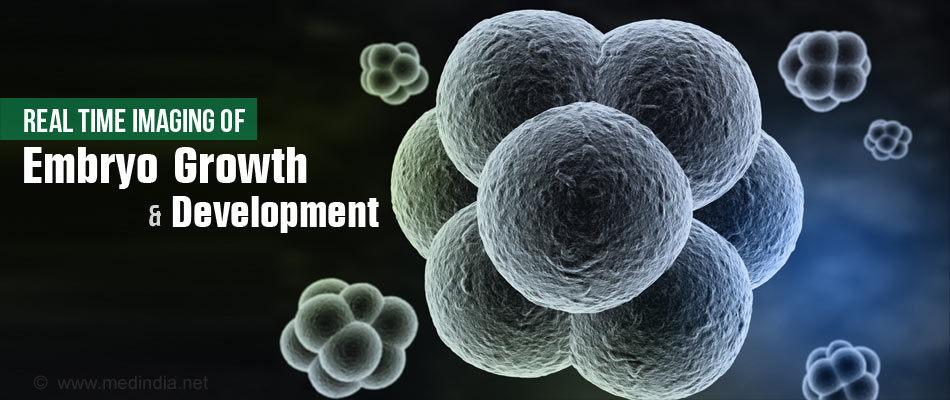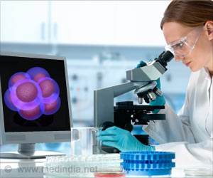
- Advancements in microscopy allow real time imaging of embryo development
- Early stage mammalian cell growth and differentiation witnessed
- Potential for improvements in In-Vitro Fertilisation (IVF) and Pre-implantation Genetic Diagnosis (PGD)
Assisted Reproductive Technologies (ART)
Infertility is a major health concern and childless couples worldwide depend on assisted reproductive techniques to bear a child. There are essentially three different types of assisted reproductive techniques.Intra-uterine Insemination (IUI): This method of assisted reproductive technique is for women with excess mucus plug in the cervix, men with low sperm count or poor motility of the sperms. The sperms are collected from the male and then inserted into the uterus of the female.
In-vitro Fertilization (IVF): Women are provided with hormone treatment to increase the production of eggs to improve chances of fertilization. The eggs are then retrieved from the woman and fertilized with the sperms collected from the male under laboratory conditions. Growing embryos that are ‘healthy’ are then inserted into the uterus of the woman.
Intra-cytoplasmic Sperm Insemination (ICSI): When the sperm count of the male is very low or if only a single sperm is available, even with motility problems, it is used to fertilize the egg from the woman by placing the sperm head in the cytoplasm of the egg.
Facts about Assisted Reproductive Technologies
- Worldwide an increasing number of childless couples depend on assisted reproductive technologies to conceive.
- ART was first used in the U.S in 1981
- In Singapore in 2013, 6000 cycles of IVF were conducted, 1000 cycles more than in 2012.
- In India,
- 5500 cycles of IVF-ICSI in 2000
- 21,500 cycles of IVF-ICSI in 2006
- 110,000 cycles of IVF-ICSI in 2011
- The numbers in India are increasing at a remarkable rate
- Success rate in the U.S
- Women 35 years or younger - 41-43%
- Women between 35 to 37 years - 33-36%
- Women between 38-40 years - 23-27%
- Women over 40 years - 13-18%
- An ICMR report on ART states that the success rate of ART is not more than 30%
"Most laboratories conduct studies on embryonic cells via invasive methods which do not keep the embryo alive. Our lab is the only one in the world imaging single cells in live mammalian embryos at the quantitative level, which allows us to observe every cell within an embryo at every stage of its development. Our findings as a result of this advanced technique have put forth a new paradigm of knowledge that would encourage more detailed microscopic analysis for future assisted reproduction procedures" said Dr Nicolas Plachta, Senior Principal Investigator of IMCB.
Highlights of the Findings of the Study
- The study shows that all cells in the developing embryo are not identical as was believed earlier.
- Cells in an embryo are differentiated and carry out different roles as they develop.
- There was a difference in binding of certain proteins in the different cells to their target genes.
- In depth understanding of the growth and development of embryos will aid in better success rates in ART.
- Embryos were previously determined if they were good enough for implantation based on morphology and growth properties, imaging techniques such as these would aid in better understanding of growth of the embryos. This could lead to rejection of embryos with growth disabilities or genetic malformations.
- Assisted Reproductive Technology (ART)
https://www.nichd.nih.gov/health/topics/infertility/conditioninfo/Pages/art.aspx - Assisted Reproductive Technology (ART)
http://www.cdc.gov/art/index.html - Assisted Reproductive Technologies
http://www.sart.org/sart_assisted_reproductive_technologies/ - Assisted Reproductive Technology
https://www.asrm.org/uploadedFiles/ASRM_Content/Resources/Patient_Resources/Fact_Sheets_and_Info_Booklets/ART.pdf - Assisted reproductive technology in India: A 3 year retrospective data analysis
http://www.ncbi.nlm.nih.gov/pmc/articles/PMC3963305/ - In Vitro Fertilization: IVF
http://americanpregnancy.org/infertility/in-vitro-fertilization/


