British scientists have successfully experimented on mice with an optical test that could detect Alzheimer’s and other neurogenerative disorders pretty early.
British scientists have successfully experimented on mice an optical test that detects Alzheimer’s and other neurogenerative disorders pretty early, even before symptoms develop.
The technique uses fluorescent markers which attach to dying cells which can be seen in the retina and give an early indication of brain cell death. The cells show up as green dots because they absorb the fluorescent dye.The research by scientists from the University College London is published in the journal, Cell Death and Disease. The findings could enable scientists to overcome the difficulty of investigating what is happening inside the brains of those with Alzheimer's.
They currently have to rely on expensive MRI scans or post-mortems.
Nerve cell death is the key event in all neurodegenerative disorders, with apoptosis and necrosis being central to both acute and chronic degenerative processes. However, until now, it has not been possible to study these dynamically and in real time. In this study, we use spectrally distinct, well-recognised fluorescent cell death markers to enable the temporal resolution and quantification of the early and late phases of apoptosis and necrosis of single nerve cells in different disease models. The tracking of single-cell death profiles in the same living eye over hours, days, weeks and months is a significant advancement on currently available techniques. We identified a numerical preponderance of late-phase versus early-phase apoptotic cells in chronic models, reinforcing the commonalities between cellular mechanisms in different disease models. We showed that MK801 effectively inhibited both apoptosis and necrosis, but our findings support the use of our technique to investigate more specific anti-apoptotic and anti-necrotic strategies with well-defined targets, with potentially greater clinical application. The optical properties of the eye provide compelling opportunities for the quantitative monitoring of disease mechanisms and dynamics in experimental neurodegeneration. Our findings also help to directly observe retinal nerve cell death in patients as an adjunct to refining diagnosis, tracking disease status and assessing therapeutic intervention, the researchers said in their article.
“In conclusion, the application of this new technology has far-reaching implications for studying intracellular processes at the single-cell level in vivo. Although the equipment we use in these studies has been customised to suit animal models, the instruments are essentially the same as those used in hospitals and clinics around the world. This raises the possibility that in the near future, clinicians may be able to assess retinal nerve cell death in vivo as a method of monitoring disease progression and treatment efficacy. Whether a single snapshot or long-term in vivo observation over many weeks is needed, the ability to visualise and track changes in cell viability offers the potential for major advances in the diagnosis and management of neurodegenerative diseases,” the scientists added.
Professor Francesca Coredeiro, lead author from University College London Institute of Ophthalmology said: "Few people realise that the retina is a direct, albeit thin, extension of the brain.
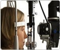
"It is entirely possible that in the future a visit to a high-street optician to check on your eyesight will also be a check on the state of your brain."
She said the research could help scientists to see how the disease is progressing by comparing retinal cell death a few weeks apart.
The first patient trials to assess the technique for the eye disease will begin later this year.
Source-Medindia
GPL
 MEDINDIA


 Email
Email


