About Careers MedBlog Contact us
Medindia LOGIN REGISTER
Advertisement

3-D Microsurgical Anatomy of Brainstem Explored by Researchers

by Himabindu Venkatakrishnan on December 2, 2014 at 2:43 PM
Font : A-A+

 3-D Microsurgical Anatomy of Brainstem Explored by Researchers

Intricate fiber dissection techniques was used in a study to provide new insights into the deep anatomy of the human brainstem, helping to define "safe entry zones" for neurosurgeons performing brainstem surgery. This was reported in a special article published in Operative Neurosurgery, a quarterly supplement to Neurosurgery, official journal of the Congress of Neurological Surgeons. These publications are published by Lippincott Williams & Wilkins, a part of Wolters Kluwer Health.

Neurosurgeons Dr. Kaan Yagmurlu and Dr. Albert L. Rhoton, Jr, of University of Florida, Gainesville, and colleagues performed fiber tract dissections, along with three-dimensional photography, to explore and document the complex anatomy of the brainstem. "We hope that the use of this knowledge will make surgical entry into the brainstem more accurate and safe," Dr. Rhoton and coauthors write.

Advertisement

Fiber Tract Dissections Guide Safe Routes for Brainstem Surgery Working under the operating microscope, the researchers performed meticulous dissections of human brainstem specimens. They used a technique called fiber tract dissection, which traces the complex course of the nerve fibers that make up the brainstem. At each step of the dissection, 3-D photographs were obtained. In some areas, a magnetic resonance imaging technique called diffusion tensor imaging was used to help in visualizing the nerve fiber tracts.

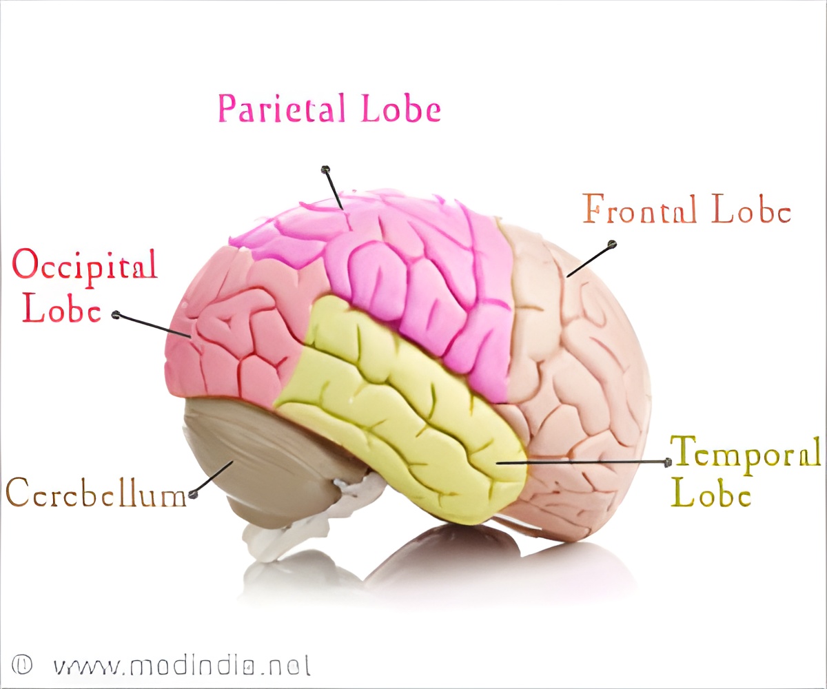
The lowermost portion of the brain is directly connected to the spinal cord. The brainstem is involved in regulating some of the body's most basic functions. In addition to mapping and illustrating the inner anatomy of the brainstem, the researchers sought to identify the best surgical approaches to specific brainstem areas.
Advertisement

In their article, Drs. Yagmurlu and Rhoton and colleagues present richly detailed descriptions and 3-D images of the fiber tract anatomy of the brainstem. They also propose a short list of "safe entry zones" for each of the three major portions of the brainstem: the midbrain, pons, and medulla.

For each area, the suggested routes include key anatomical landmarks for the neurosurgeon to follow. The fundamental principle is to pass through as little normal tissue as possible, while avoiding critical areas and tracts.

"The safest approach for lesions located below the surface is usually the shorter and most direct route," the researchers write. However, for some parts of the brainstem, a more circuitous approach is needed to reach the target while protecting essential functional areas.

Although previous studies have used fiber dissection techniques to examine safe entry zones to the brainstem, most have focused on the surface anatomy. The new study is the first to use these techniques to define and document the "complex internal architecture" of the brainstem.

Drs. Yagmurlu and Rhoton and coauthors note that their dissection study cannot account for the infinite variations in anatomy of the human brainstem--the use of neurophysiological monitoring and mapping remains crucial during surgery. However, by providing unprecedented descriptions and 3-D images of the inner anatomy of the brainstem, the researchers hope their work will help neurosurgeons in planning the best approach to microsurgical procedures for gliomas (brain cancers) and blood vessel malformations in each area of the brainstem.

Source: Eurekalert
Advertisement

Advertisement
Advertisement

Latest Research News

 Brain Region Linking Short-term to Long-term Memory Discovered
The model for memory consolidation claims that the hippocampus forms new memories and, as time goes on, trains the cortex to store enduring memories.
What Are the Effects of T Cells on Blood Pressure and Inflammation?
A new study explored the link between T immune cells in ill patients and mortality risk.
How Does a New Procedure Help Patients Avoid Leg Amputation?
Limb savage procedure benefits patients with severe vascular disease who are at risk for amputation of their limbs.
Omega-3 Can Save Alzheimer's Patients from Vision Loss
Does omega-3 help Alzheimer's patients? A new form of omega-3 helped restore specific markers of eye health in mice bred with aspects of early-onset Alzheimer's disease.
Why Is Asthma Linked to Increased Risk of Osteoarthritis?
Drugs used to inhibit the physiological responses for allergic reactions lessen osteoarthritis risk, revealed research.
View All
This site uses cookies to deliver our services.By using our site, you acknowledge that you have read and understand our Cookie Policy, Privacy Policy, and our Terms of Use  Ok, Got it. Close
×

3-D Microsurgical Anatomy of Brainstem Explored by Researchers Personalised Printable Document (PDF)

Please complete this form and we'll send you a personalised information that is requested

You may use this for your own reference or forward it to your friends.

Please use the information prudently. If you are not a medical doctor please remember to consult your healthcare provider as this information is not a substitute for professional advice.

Name *

Email Address *

Country *

Areas of Interests