The process of tissue formation in the human heart has been unraveled in research on frog embryos.
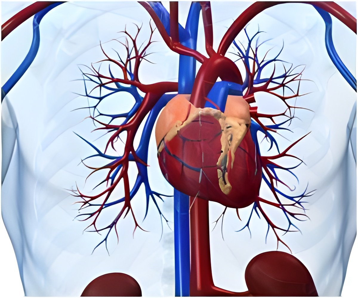
Jean-Pierre Saint-Jeannet, a developmental biologist at Penn's School of Veterinary Medicine, along with Young-Hoon Lee of South Korea's Chonbuk National University were behind the finding.
"In the frog, we were expecting to find something that was in between fish and higher vertebrates, but that's not the case at all," said Saint-Jeannet.
"It turns out that cardiac neural crest cells do not contribute to the outflow tract septum, they stop their migration before entering the outflow tract. The blood separation comes from an entirely different part of the embryo, known as the 'second heart field'," he said.
Knowing these paths, and the biological signals that govern them, could have implications for human health.
"There are a number of pathologies in humans that have been associated with abnormal deployment of the cardiac neural crest, such as DiGeorge Syndrome," said Saint-Jeannet.
Advertisement
"Xenopus could be a great model to study the signals that cause those cells to migrate into the outflow tract of the heart.
Advertisement
The study will be published in the May 15 edition of the journal Development.
Source-ANI

