Ileal conduit is the most commonly done diversion procedure, whereas 'Cutaneous Ureterostomy' is less common and has been used in infants and children when they have congenital defects of the bladder.
One or both ureters are detached from the bladder. They are brought through a new opening made on the abdominal wall or into a section of small intestine and the intestine is brought onto the abdominal wall. The urine collects into the stoma pouch that is fitted snugly against the abdomen with the help of a belt.
For creating an ileal conduit, the ileum section of small intestine is used to create the pouch.
The urinary tract comprises the kidneys, ureters, the bladder and the urethra. The kidneys filter the circulating blood to remove unwanted materials. The two kidneys present in the body filter about 120 to 150 liters of blood every day and produce up to 2 liters of urine per day.
The ureters are thin tubes coming out of each kidney and carry the urine to the urinary bladder. Urine is stored in the bladder until it is full and the brain receives a signal to void.
The bladder is a hollow organ that is located in the lower section of the abdomen and has a capacity of about 400 mls. The bladder essentially has two important functions:
- It helps to store urine.
- It empties the urine when it is full. A child learns to void when the bladder is full though training by the mother.
Urine is voided from the bladder through a tube called urethra, located at the bottom of the bladder. When ureterostomy is done, the bladder and the urethra are bypassed.
- Cancers of bladder where bladder needs to be removed
- Neurogenic bladder where the kidney function is compromised
- Irreparable loss of bladder due to injury
Cancer of Bladder
The usual cancer of the bladder is called transitional cell carcinoma and if the cancer is superficial, it indicates a good prognosis and the chances of survival are high. However if it is deep and invasive, then the survival rate declines and hence removal of bladder and a urinary diversion or an artificial bladder may need to be created.
Neurogenic Bladder
Neurogenic bladders are abnormal bladders where either the sensation to empty is lacking or the whole voiding mechanism is un-coordinated. Some of these bladder contracts or becomes overactive even when resting and not full for voiding.
The filling and voiding mechanism of the bladder are controlled by the brain through nerves that pass down from the brain via the spinal cord and supply the bladder. One set of nerves takes the sensation of bladder fullness to the brain and another set from the brain commands the bladder to empty. When these nerves are either deficient or get damaged it leads to the condition called neurogenic bladder.
Neurogenic bladder cannot only give symptoms of urgency and incontinence but also can damage the kidney due to high pressures within the bladder that get transmitted to the kidneys. Urinary diversion procedures in such situations are done to protect the kidneys from these abnormal bladders.
Recently at Wake Forest Research Centre in the USA, Neo bladder from 'Tissue Engineering' has been created to replace such diseased bladders.
What Causes Neurogenic Bladder?
There are a number of causes that lead to 'Neurogenic bladder'. Some babies are born with problems of the spinal cord, such as spina bifida and these lead to abnormal bladder. The average incidence of spina bifida is 1 case per 1000 births in most countries. Other conditions that lead to neurogenic bladder include:
- Spinal trauma
- Spinal surgery for disc
- Diabetes
- Multiple sclerosis
- Major pelvic surgery
- Cerebral palsy
- Parkinson's disease
- Cerebral stroke
- Urinary bladder with fistula's
- Heavy metal poisoning
- Bladder cancer or tumor
Opening Brought Out to the Skin
- Ileal conduit
- Cutaneous
- Continent Urinary Diversion
Opening Diverted to the Bowel or Intestine
Uretero-colic Diversion
To the Urethra
Orthotopic Neo bladder
Opening Brought Out to the Skin
Urostomy stoma can be done in two types of procedures - ileal conduit and cutaneous stoma.
Ileal Conduit: A short segment of the small intestine is taken away, while the remaining of the small intestine is reconnected to function normally. The removed segment is placed in such a way that it connects kidneys to a stoma on the skin surface via the ureters.
A temporary fine tube called urostomy stents are inserted into each kidney via the ureters to allow free drainage of the urine from the kidney into the small intestine conduit or stoma bag.
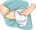
The urine now travels directly from the kidneys through the newly formed passageway through the stoma to an external collecting pouch. The pouch is fixed externally on the skin and can be drained into a toilet without disconnecting it from the stoma.
Cutaneous Ureterostomy: The ureters are detached from the bladder and the opening is brought to the surface of the abdomen. A stoma, or opening, is created at the point and an external pouch is fitted at the stoma.
Cutaneous ureterostomy can be done in four ways. Single ureterostomy is a procedure in which only one ureter is brought to the surface of the abdomen. When the two ureters are brought to the surface of the abdomen, one on each side of the abdomen, the procedure is called bilateral ureterostomy.
Double-barrel ureterostomy involves bringing together both the ureters to the same side of the abdominal surface, through two different stomas.
The pouch needs to be emptied when it is filled to about one-third to one-half. During night, a piece of flexible tubing can be attached to the draining valve to empty into a bigger pouch, so that the patient is not disturbed during their sleep.
The Ureterostomy Stoma is moist and red and produces mucus as it is a part of the bowel that is modified. It does not have any sensation as there are no nerve endings, but has rich blood supply. The stoma may seem to move sometimes, but this is not a cause of concern. It is in response to the normal contractile movement of the bowel that directs the urine towards the urostomy pouch.
The Urinary Pouch or bag called a urostomy bag is made of a plastic substance and it is fitted on to the ileal conduit or cutaneous ureterostomy. The pouch system consists of two parts.
- Wafer - a barrier of the pouch that sticks to the skin.
- A disposable bag or pouch that attaches to the wafer.
Sometimes the two units are fused into one. The barrier keeps away the urine from the skin and is designed to be skin-friendly. The pouch is connected to the barrier and has a draining valve at the bottom. The draining valve helps the patient to empty the urine into a toilet without removing the pouch from the stoma.
A one-piece pouch comprises of a pouch with the adhesive directly on to it, and can be left on the skin for 1 to 3 days before it is changed. A two-piece pouch system has a flange for the skin barrier, which is fitted to the abdomen around the stoma, and the pouch is fitted into the flange. This type of pouch can be retained for about 3 to 5 days.
The urostomy pouch is available in shops selling surgical goods, under several brand names. All pouches are made of water-proof and skin friendly materials and are odor-proof. The valve that collects the urine is usually a one-way valve that prevents the urine in the pouch from flowing back to the stoma. The valve that drains out urine is fitted at the bottom of the pouch.
Continent Urinary Diversion Procedures
Special diversion procedures called continent urinary diversion can be performed but these are more complicated to perform and sometimes can have higher early surgical complication rate. In this procedure, the surgeon creates an internal reservoir from a section of the intestine, and connects the ureters into this reservoir once it is ready. The patient can drain the urine collected in the reservoir pouch using a small tube. The urine will need to be drained 4 to 5 times a day with these catheters. This procedure does not require wearing an external pouch to drain the urine.
Continent urinary diversion can be done in two ways:
- Continent cutaneous reservoir: A reservoir is created under the skin surgically and then is connected to a stoma on the surface of the skin. The patient can insert a catheter through the stoma into the reservoir to drain the collected urine several times in a day. The stoma is usually less than 2 cms wide and can be attached to the belly button so that it remains hidden. There is no need of an external pouch for this type of urostomy as mentioned previously.
- Bladder substitute or Orthotopic Neo bladder: An internal reservoir is created by the surgeon and is connected to the ureters at one end and to the urethra at the other end. The urine collected in the reservoir through the ureters can be drained via the urethra in a more natural process. However, the functioning of such a bladder is not similar to the natural one. Sometimes a catheter must be inserted through the urethra into the reservoir to drain out the urine collected or there may be chances of urinary incontinence or infection. But this is the preferred method for many patients as they retain the normal external anatomy.
Uretero-colic Diversion or Ureterosigmoidostomy
This is an internal diversion to where the ureters are diverted to the large intestine such as sigmoid colon and urine gets mixed up with feces. This procedure does not require care of the external stoma and is sometimes the preferred procedure in children born with bladder abnormality such as extrophy of the bladder or when there is a cancer of the bladder such as rhabadomyosarcoma. It is also the preferred method in a less affordable patient, where the costs of the external appliances need to be avoided. The disadvantage of the procedure is that there is a risk of infections of the kidneys and salt imbalance due to absorption from the intestine.
Preparatory Diagnostic Tests for Urinary Diversion Procedures
Patients who are advised to go for urinary diversion procedures will have to undergo diagnostic procedures:
- Blood tests like complete blood count and electrolytes
- Kidney or renal function tests - levels of blood urea, nitrogen and creatinine.
- Imaging studies of kidney, ureters and renal pelvic area like:
- Intravenous urogram or pyelogram (IVU/IVP): A test that involves injecting a contrast dye into a vein and is followed through its time course of excretion through the kidneys, ureters and bladder.
- Retrograde pyelogram: X-ray focusing on the region of the kidney and ureters, where the urine collects.
- Antegrade Nephrostogram CT-scan: Multiple X-ray images are collected to form a two-dimensional cross-sectional image of the kidneys and the ureters.
- MRI with intravenous gadolinium: Imaging studies of the internal soft tissues of the body.
Postoperative Care After Urinary Diversion Procedures
After the surgical procedure, it is very important to take care of the stoma. This is because, the stoma is directly connected to the kidney through the ureters and all these organs are exposed to infections.
There are specially trained nurses and para-medical staff who can train a person in how to use the urostomy bag and work as a support staff. There are also support groups that can help.
A WOC nurse (Wound, Ostomy and Continence nurse) advices and helps the patients to take care of the wound, stoma and the pouch. Entero-stomal therapists counsel the patients regarding ostomy care and rehabilitation.
Postoperative Care after Urinary Diversion Procedures - Taking care of the stoma:
Any continent or non-continent stoma requires basic daily care to keep the skin surrounding the stoma healthy and clean. Cleaning can be done in the following ways:
- Extra mucus must be wiped away as soon as it is spotted.
- Rinse the stoma with clean water.
- Wash the stoma and the surrounding skin with mild soap and water.
- Dry the stoma completely.
Taking Care of the Pouch:
- Rinse and clean the pouch daily.
- Change the pouch every 5 to 7 days.
- While changing the pouch, the skin around the stoma must be wiped with a wet towel.
- Before attaching the new pouch, the skin must be completely dry.
- Skin around the stoma may be irritated with constant flow of urine. Protective skin wipes or an ostomy powder can be used in such cases.
Care for Continent Cutaneous Reservoir:
- Learn how to insert a catheter through the stoma or urethra to drain the internal reservoir.
- Drain the internal reservoir every couple of hours during the first few weeks after surgery. There after the frequency may reduce.
General Care after Urinary Diversion:
- Avoid lifting heavy objects and doing strenuous physical activities.
- Talk to your enterostomy specialist and employer if adjustments have to be made in job responsibilities.
- Avoid contact sports such as soccer, football and martial arts.
- Normal diet can be resumed after surgery. However, some food items, such as seafood, may cause strong odor in urine. If this is a concern for a patient, such food must be avoided.
- Urine testing must be done at frequent intervals to identify any infection and treatment must be given immediately.
Bacterial infection is one of the post-operative risks of urinary diversion ureterostomy. Symptoms like fever, milky urine, strong-smelling urine and urine containing excessive mucus, must be looked into and further investigations must be carried out.