Medindia
Medindia LOGIN REGISTER
 Osteotomy

Osteotomy

Last Updated on Jul 28, 2017
Font : A-A+

What is Osteotomy?

Osteotomy is a surgical procedure that involves cutting the bone in order to shorten or lengthen it or to realign it with respect to another adjacent bone.

What are the Types of Osteotomy?

There are several types of osteotomy depending on the part on which the surgery is performed:

  • Knee Osteotomy: This type of osteotomy is used for realigning the knee joint so that the weight-bearing function is shifted from the damaged side to the healthy side of the knee, in order to prevent the arthritic surfaces from rubbing against each other. In a knee osteotomy, the shinbone (tibia) or thighbone (femur) is cut and reshaped so as to relieve pressure on the knee joint. This is usually carried out in case of early stages of osteoarthritis, where the damage has occurred on one side of the knee joint.
  • Hip Osteotomy: This is also known as femoral osteotomy and is used to treat hip dysplasia. It involves adjustments made to the ball of the thighbone (femur head) and the socket (acetabulum) of the hip bone. The bones are cut and reshaped to realign the load-bearing surfaces of the “ball-and-socket” joint.
  • Jaw Osteotomy: Osteotomy of the jaw is carried out to realign the lower jaw (mandible) or upper jaw (maxilla). The surgery is done inside the oral cavity. Sometimes dental braces may need to be worn before and after the surgery to realign the teeth with reference to the newly repositioned jaw.
  • Chin Osteotomy: This type of osteotomy is carried out most often to extend the vertical length of the chin, which in this case is shorter than normal.

Why is Osteotomy Done?

An osteotomy is done for the following major reasons:

  • Weight Distribution: The surgery enables transfer of weight from the damaged area to the healthy area, thereby providing optimal weight distribution.
  • Alignment of the Joint: In case of knee osteotomy, the surgery helps to align the affected leg with reference to the healthy leg so that bowlegged alignment is corrected. In case of hip or femoral osteotomy, the surgery helps to align the hip joint and articulate it properly with the head of the thighbone so that optimal weight distribution occurs.
  • Conservation of the Joint: The surgery helps to increase the lifespan of the joint and delays the need for an artificial prosthesis. For example, in case of a knee osteotomy, the surgery can delay the need for a total knee replacement by up to 10 years.
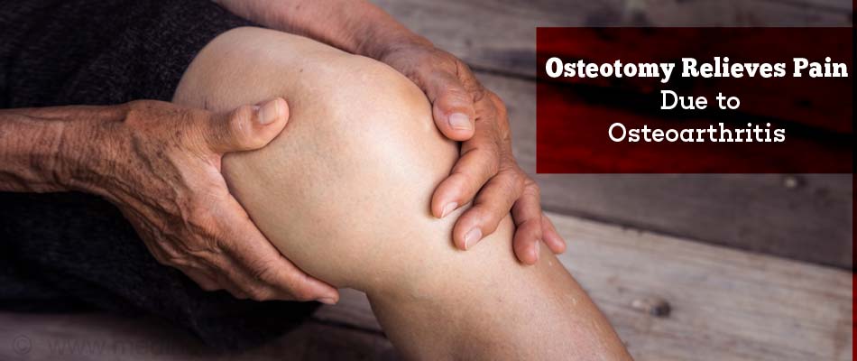
  • Pain Relief: The surgery relieves pain arising from osteoarthritis by adjusting weight distribution in the joint.
Osteotomy Relieves Pain Due to Osteoarthritis

How do you Prepare Before the Osteotomy?

Routine Tests: For all patients who undergo an elective surgery (which is more planned), routine tests to evaluate the health status of the patient are required and include the following:

  • Blood tests like hemoglobin levels, blood group, liver and kidney function tests
  • Urine tests
  • ECG to study the electrical activity of the heart
  • Chest X-ray
  • Detailed assessment of heart function (senior patients)

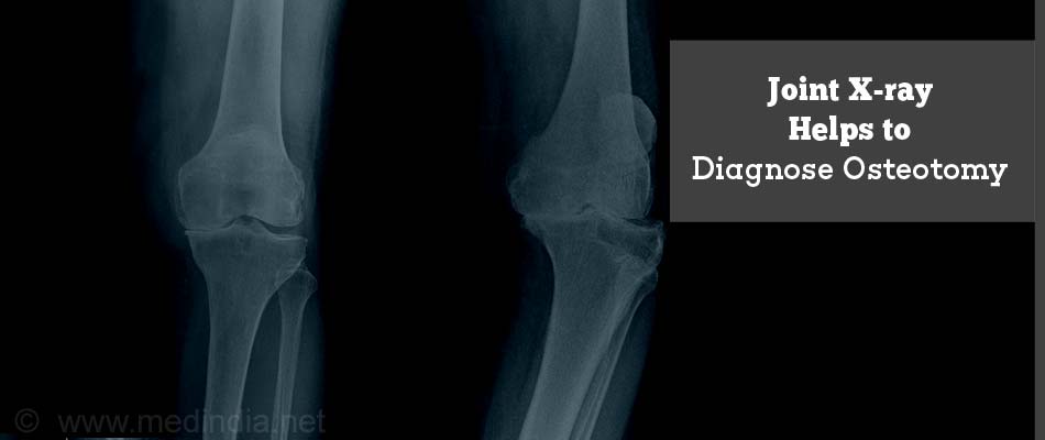
X-Ray and CT Scan: X-rays and computed tomography (CT) scans may be used to take images of the joint so that the area of the surgery can be carefully mapped. Sometimes, computer modeling is also used to do the mapping. This enables the surgeon to exactly calculate the size, dimensions and angles of the wedge that needs to be excised.

For a CT scan, you will be asked to lie down still on a special table for a few minutes. A doughnut-shaped instrument will pass over the part to be screened, and images will be taken. A contrast agent may be injected into your vein; therefore you should inform the radiologist if you suffer from any allergies.

Joint X-ray Helps to Diagnose Osteotomy

Pre-operative Check-ups and Precautions: Routine tests as indicated above are ordered a few days before the surgery. Admission is required a day or two before the surgery. If you are on blood thinners or aspirin, these should be stopped a few days before the procedure.

Fasting on the Night Before Surgery: Overnight fasting is required and occasionally intravenous (IV) fluid may be required to keep you well hydrated. Sedation is sometimes required for good overnight sleep before the surgery.

Type of Anesthesia: Osteotomy can be done either under general anesthesia (GA) (which will put you to deep sleep) or spinal anesthesia (you will be awake but your body will be numb from the waist down).

What Happens During the Surgery?

Shift from the Room or Ward to the Waiting Area in the Operating Room: An hour or two before the surgery, you will be shifted to the operating room waiting area on a trolley. Once the surgical room is ready, you will be shifted to the operating room.

Shift to the Operating Room: The ambiance in the operating room can sometimes be very daunting and a small amount of sedation can help overcome your nervousness. From the trolley, you will be shifted on to the operating table.

Anesthesia Before Surgery: If the anesthetist decides to administer GA, you will be injected with drugs through an IV line and made to inhale some gases through a mask that will put you into a deep sleep for anesthesia. Once you are in deep sleep, a tube will be inserted into your mouth and windpipe to administer the anesthesia gases to overcome pain and keep you comfortable. If on the other hand the anesthetist decides to administer spinal anesthesia, you will be given an injection into the lumbar region of your backbone (vertebral column), which will numb your body from the waist down.

Surgical Procedure: The osteotomy procedure is discussed, taking tibial osteotomy as an example. Tibial osteotomy, also known as “high tibial osteotomy” was first performed in Europe in the 1950s. This procedure is carried out as a treatment for knee osteoarthritis, in order to correct a bowlegged alignment that can put pressure on the inner (medial) side of the knee. The surgery generally lasts between 1-2 hours.

  • The leg is first sterilized with antibacterial solution and draped.
  • During the procedure, the surgeon makes a 4-5 inch incision at the front of the knee, below the kneecap and extending below the top of the shinbone (tibia).
  • The surgeon uses guidewires to help cut out a wedge of bone from the outer (lateral) side of the shinbone on the healthy side of the knee joint. The guidewires usually outline a triangular form in the shinbone.
  • An oscillating saw cuts out the wedge from underneath the knee, below the healthy cartilage, which is left intact.
  • The wedge of bone is removed and the gap is “closed” by bringing together the bones in order to fill the space created by removing the wedge.
  • A plate and screws will be inserted to hold the bones in place till the osteotomy heals.
  • The wound is closed by stitching up the different layers of tissue in the knee using absorbable sutures. This is the most common type of osteotomy procedure, and is called a “closing wedge osteotomy”. This procedure realigns the joint so that the bowleg is corrected and the pressure is shifted to the healthier side, thereby easing the pressure on the damaged side.
  • Sometimes, instead of “closing” the wedge, it is “opened up” and a bone graft is inserted to facilitate healing. This procedure is called “opening wedge osteotomy”.
  • Another type of osteotomy, called Fulkerson osteotomy is done to realign the tibial tuberosity (lower bony attachment of the patellar tendon on the shinbone).
  • A similar type of osteotomy is done on the thighbone (femur) to correct knock-kneed alignment.

What Happens after the Osteotomy?

Waking up from Anesthesia: Once the surgery is over you will wake up and the tube down the wind pipe will be removed. You will be asked to open your eyes before the tube is removed. Once fully awake, you will be shifted on the trolley and taken to the recovery room.

Recovery after Osteotomy

Recovery Room: In the recovery room, your vitals will be monitored and observed for an hour or two before shifting you to the room or a ward. Injections for relief of pain will be administered to ensure proper sleep, until oral intake is resumed. Antibiotics will be given to prevent infectionS.

Post-operative Recovery: You will remain in the hospital for a few days following the procedure. Normally, on the first day, you will not be allowed much to drink or eat. Once the bowels start recovering, you will be given fluids and a light diet. This may take one to three days.

Change of dressing will be done as required. The IV lines for fluids and drugs will continue for a few days till you start eating normally.

Once you are stable and have resumed normal diet, you will be discharged with detailed instructions to be strictly followed to ensure optimal recovery. Oral pain killers may be prescribed to be taken as needed.

You may be advised to attend a check-up a week after discharge to check how the wound is healing.

Other aspects of post-operative recovery, which is specific to osteotomy include the following:

  • Weight-bearing on the Operated Leg: After the surgery, you will need to use crutches for several weeks to prevent pressure on the operated leg. The surgeon may put a brace or a cast on your knee to help the healing process and will also instruct when you can resume weight bearing on the affected leg and start rehabilitation exercises.
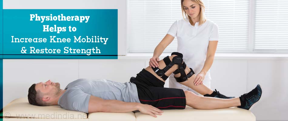
  • Physiotherapy: A physiotherapist will show you some exercises that you can do during the rehabilitation period. These exercises will increase the mobility of the knee and help to restore strength.
Physiotherapy Helps to Increase Knee Mobility & Restore Strength
  • Resuming Work: You will be able to return to work and resume your full activities in 3-6 months after the surgery.

What are the Risks & Complications of Osteotomy?

As with any type of surgical procedure, there are risks involved with osteotomy. However, the surgeon will try to minimize the chances of potential complications. The most common complications are highlighted below:

  • Infections
  • Blood clots (thrombosis)
  • Injuries to blood vessels and nerves during surgery
  • Stiffness of the knee joint
  • Failure to heal - It should be noted that a second surgery may be required if the osteotomy fails to heal properly.

Recommended Readings

Post a Comment

Comments should be on the topic and should not be abusive. The editorial team reserves the right to review and moderate the comments posted on the site.

Get Health and Wellness Secrets from Our Engaging eBooks
Advertisement
Surgical Procedures A-Z
A B C D E F G H I J K L M N O P Q R S T U V W X Y Z
Advertisement
OSTEOTOMY - Related News Why Is Osteoporosis So Misunderstood and Ignored?
Say Goodbye to Injections, Hello to Oral Relief With Robotic Pills for Osteoporosis
New, Quick Blood Test Helps Diagnose Osteoporosis Risk
New Potential Osteoporosis Treatment Identified
World Osteoporosis Day: Let's Build Better Bones
'Build Better Bones' Platform Expands Multilingual Osteoporosis Support
Osteoporosis Prevalence: India Leads Worldwide
Osteoporosis in Youth: The Silent Consequence of Modern Lifestyles
Female Ginseng's Role in Osteoporosis Therapy
Living Near Green Spaces Tied to Stronger Bones and Lower Osteoporosis Risk
More News
What's New on Medindia
World Malaria Day: Ensuring Equitable Access
Test Your Knowledge On Sunscreen
Teen Mental Health Awareness: Signs That Every Parent Should Know
View all
Stay Connected
Available on the Android Market Available on the App Store
This site uses cookies to deliver our services.By using our site, you acknowledge that you have read and understand our Cookie Policy, Privacy Policy, and our Terms of Use  Ok, Got it. Close