Diagnosis
Microscopic analysis of the peripheral blood and bone marrow along with cytogenetic evaluation confirms the diagnosis of CML.
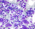
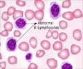
1. A microscopic analysis of the peripheral blood of a CML patient will reveal the following -
- Elevated number of granulocyte cells especially basophils and eosinophils. This feature helps to differentiate CML from a leukemoid reaction.
- The blood test will also typically reveal immature myeloid cells.
2. A bone marrow biopsy is often performed as part of CML evaluation. During the biopsy, a needle is inserted into the hip bone to draw out a small amount of bone marrow cells. This is then subjected to a microscopic analysis. This will help the health care expert to understand the stage of the disease and to plan. Treatment accordingly other tests need to be carried out to estimate the extent to which the disease has spread.
3.The ultimate test for CML is the detection of the Phildelphia (Ph) chromosome.This characteristic cytogenetic abnormality can be identified by -
- Routine karyotyping
- Fluorescent In Situ Hybridisation (FISH)
- Polymerase Chain Reaction (PCR) technique
There is debate over the ‘Ph-negative’ CML in which the Ph chromosome cannot be detected. Many of these patients harbor other chromosomal abnormalities of a complex nature which masks the (9;22) translocation.In some patients the karyotyping may be normal but the translocation may be evident when FISH or a RT-PCR is carried out.These patients without detectable molecular evidence of the translocation may be better classified as suffering from undifferentiated myelodysplastic or myeloproliferative disorder.In these patients,the clinical course tends to be different from patients with CML.


