- Tracheal cancer (cancer of the windpipe). Reviewed Mar 31, 2016. Accessed July 13, 2018. Cited July 13, 2018. - (https://www.macmillan.org.uk/information-and-support/trachea-windpipe- cancer)
- Napieralska A, Miszczyk L, Blamek S. Tracheal cancer – treatment results, prognostic factors and incidence of other neoplasms. Radiology and Oncology. 2016;50(4):409-417 - (doi:10.1515/raon-2016-0046. - )
- Tracheal cancer. Patient Education. Accessed July 13, 2018. Cited July 13, 2018. - (https://www.cooperhealth.org/sites/default/files/pdfs/Tracheal %20Cancer.pdf)
- Compeau CG, Keshavjee S. Management of tracheal neoplasms. The Oncol. 1996;Vol1(6):347-353. - (http://theoncologist.alphamedpress.org/content/1/6/347.full.html)
- Nouraei SM et al. Management and prognosis of primary tracheal cancer: A national analysis. Laryngoscope. 2014;124:145-150. - (http://www.sbccp.org.br/arquivos/LG2014-01/lary24123.pdf)
- Kumar R et al. Tracheal tumor: A diagnostic and therapeutic dilemma. 2015;Vol.4(2):129-133. - (http://www.ccij-online.org/article.asp?issn=2278- 0513;year=2015;volume=4;issue=2;spage=129;epage=133;aulast=Kumar)
- Hetna³ M, Kielaszek-Æmiel A, Wolanin M, et al. Tracheal cancer: Role of radiation therapy. Reports of Practical Oncology and Radiotherapy. 2010;15(5):113-118 - (doi:10.1016/j.rpor.2010.08.005 - )
What is Tracheal Cancer?
Cancer of the trachea affects the tube connecting the mouth and nose to the lungs. It is a rare form of cancer without a specific known cause. Tracheal cancers are classified as primary and secondary carcinomas. Primary cancer arises from the trachea while secondary cancer is due to cancer that has spread to the trachea from another site.
- Almost 75% of primary tracheal cancers are either squamous cell carcinoma (SCC) (most common) or adenoid cystic carcinoma (ACC). Adenoid cystic carcinomas are found in equal measure in men and women between the ages of 40 and 50 and are responsible for ~1% of head and neck cancers. The rate of progression is slow in ACC. Men between the ages of 60 and 70 are invariably affected with SCC.
- Secondary tracheal carcinomas are tumors that spread to the trachea from the lungs, esophagus, thyroid, or larynx.
Facts and Statistics about Tracheal Cancer
- Primary tracheal carcinomas account for 0.01% - 0.4% of all malignant cancers.
- Nearly 90% of primary tracheal tumors in adults are malignant.
- The overall survival is better in patients with ACC (10% to 89.4%) than with SCC (7% to 46%).
What are the Stages of Tracheal Cancer?
Tracheal cancer is rare and does not have a standardized staging or grading system.
In general, tracheal tumors are clinically staged as
- Early or local – Cancer that is limited to the trachea.
- Locally advanced – Cancer that has invaded into nearby sites of the body.
- Metastatic or advanced– Cancer that has spread to distant sites, such as the liver, lungs, or bones.
Grading of tracheal cancer
Grading of tracheal cancer is done by looking at the appearance of the tumor cells under the microscope. Based on their appearance, the tumor may be graded as
- Low-grade - Cancer cells resemble normal cells of the organ
- High-grade – Cancer cells don’t look like normal cells of the organ and look abnormal
A low-grade tumour grows more slowly and be less likely to spread to other areas than a high-grade tumour.
What are the Causes of Tracheal Cancer?
There is no clear cause of tracheal cancer. Smoking is considered to cause SCC. In general, it is not clear how primary tracheal cancer develops. Tracheal cancers can affect humans at any age.
What are the Symptoms & Signs of Tracheal Cancer?
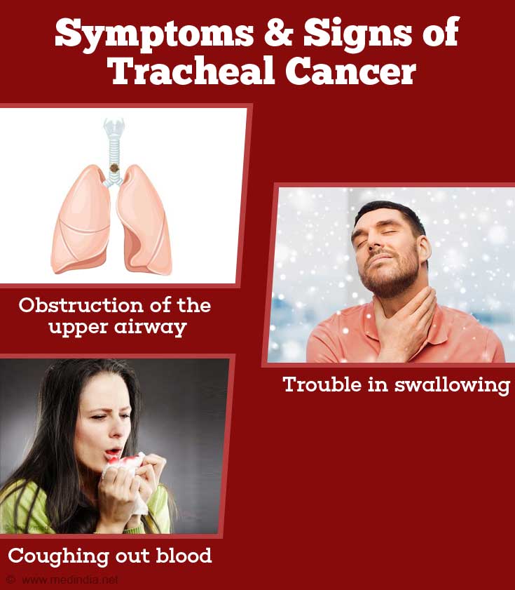
Tracheal cancer is characterized by different symptoms as listed below:
- Obstruction of the upper airways – primary symptom
- Trouble in swallowing (dysphagia)
- Dry cough
- Coughing out blood or blood-stained mucus (hemoptysis)
- Hoarse vocal sounds
- Recurring fever, chest congestion and chills
- Breathing noisily and wheeze
- Loss of breath, difficulty in breathing (dyspnea)

What are the Consequences of Tracheal Cancer?
Tracheal cancer is very rare and complications arise following surgery. These complications include pneumonia, irregularities in wound healing, and inflammation in the region surrounding the trachea. Other complications arising from local tumor invasions, are damage and destruction of the trachea and tracheal narrowing or stenosis, and bleeding from the tumor.
How do you Diagnose Tracheal Cancer?
You should visit your family physician when you begin to experience any troubling symptoms. Your doctor will take your medical history and conduct a physical examination. If cancer is suspected, you will be recommended to a specialist who will perform additional tests to confirm the diagnosis.
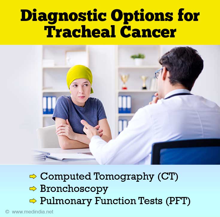
- Computed tomography (CT) scans and chest tomography help to diagnose and identify the stage of tracheal cancer according to the spread and localization of the tumor. Based on the cancer stage, it is easy to decide on an appropriate treatment strategy. Three-dimensional helical CT scans help to plan the surgical removal of tumors because they provide information on the location and length of tracheal tumors. Tumor invasion into the esophagus can be identified using contrast esophagography combined with CT.
- Bronchoscopy, an important technique for staging and diagnosis, helps to understand how and where tracheal tumors have spread. Tissue biopsies are obtained for further diagnostic investigation. The bronchoscope consists of a camera and a light attached to the end of a flexible tube (flexible bronchoscopy), and it is introduced through the nose or mouth, into the body’s air passages (trachea and bronchi). When the tube is rigid, it is called rigid bronchoscopy and this is used in an operating room to confirm the CT scan diagnosis of upper airway obstruction. The bronchoscope is sent past the obstruction and the trachea is then widened using endoscopic measures.
- Pulmonary function tests or lung function tests examine the condition of the lungs to assess how well they are performing. Obstruction of the upper airways indicates possible tracheal cancer.

How Can you Treat Tracheal Cancer?
It is very important to diagnose tracheal cancer at its earliest. The trouble is that in most cases, obstruction of the upper airways to the lungs is often misdiagnosed as asthma. And so, when the individual is finally diagnosed with tracheal cancer, it is invariably in the advanced stage resulting in low survival rates. There is also limited knowledge of treatment options.
In general, the treatment options for tracheal cancer, include surgery, endoluminal brachytherapy, radiation therapy, chemotherapy, and interventional endoscopy.

Surgery may not be required for individuals with primary low grade tracheal cancer (SCC variety) that is malignant with only local invasion.
In other cases, surgery, in combination with chemotherapy or radiotherapy are performed to effectively remove the tumor.
In advanced inoperable tracheal cancer, radiotherapy alone may be given to give relief from breathing symptoms (palliative) and keep the patient comfortable, not aimed at curing.
Surgery: There are different surgical techniques used for tracheal cancer, such as laryngotracheal resection, sleeve trachea resection, resection and primary reconstruction, and immediate tracheal reconstruction.
Surgery involves removing the portion of the trachea containing the tumor and joining the ends. The trachea is shortened and so you have to be careful when moving your head. You will be given food using a drip, and excess blood and fluids are drained out to avoid contact with, and infection of the operated region.
Brachytherapy: A tube with radioactivity is inserted into the trachea near the tumor in order to shrink it. This method helps to reduce complications that arise from external radiotherapy.
Radiotherapy: Radiation (median dose of 40 to 60 Gray) is normally administered following surgery (adjuvant treatment). In some cases, where surgery cannot be performed, radiation is the primary form of treatment.Radiotherapy may be associated with side effects such as difficulty in swallowing and indigestion
Endobronchial stents: Silicone tubes are inserted into the upper airway obstruction to ease airflow.
Chemotherapy: Drugs, such as carboplatin, cisplatin, and paclitaxel, are used in combination with radiation and surgery. Chemotherapy along with radiation is recommended if surgery is not performed.
Endotracheal debridement: Individual procedures, such as rigid bronchoscopy with suction and forceps to remove tissue biopsies, are performed. This helps to clear tracheal tumors. Laser therapy, cryotherapy, and electrocoagulation are used individually or in combination to remove tracheal tumors.


