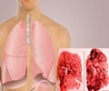
Scientists at Washington University School of Medicine in St. Louis say that a noninvasive approach towards assessing lung inflammation could hasten the process of developing drugs for cystic fibrosis and pneumonia. Positron emission tomography (PET) scans were used to monitor artificially induced inflammation in the lungs of healthy volunteers.
"Until now, when we wanted to assess whether a new drug decreased lung inflammation, the options for specifically measuring active inflammation were not pleasant," says lead author Delphine Chen, M.D., chief resident in nuclear medicine at the medical school's Mallinckrodt Institute of Radiology. "We could perform a bronchoscopy and gather samples directly from the breathing passages, or we could have patients inhale a saline solution and cough it back up." Researchers say that the process of monitoring each step in the inflammatory process will help in formulating drugs to stop the same. Their findings are published in he Journal of Applied Physiology. "Full-scale clinical trials are costly in terms of both time and dollars spent, and right now it's very difficult to find intermediate steps that allow us to build confidence in a drug's effectiveness before taking that plunge," said senior author Daniel P. Schuster, M.D., professor of medicine and of radiology. He added that drugs could now be tested in smaller groups of volunteers. "If the drug passes those tests, then you can say, okay, let's see in a full-scale trial if the drug actually has an impact on some important patient-centered outcome like mortality or disease progression," he added. Main Article: Chen DL, Rosenbluth DB, Mintun MA, Schuster DP. FDG-PET imaging of pulmonary inflammation in healthy volunteers after airway instillation of endotoxin. The Journal of Applied Physiology, online publication.PET Scans Help In Monitoring Progress of Lung Inflammation
Recommended Readings
Advertisement


