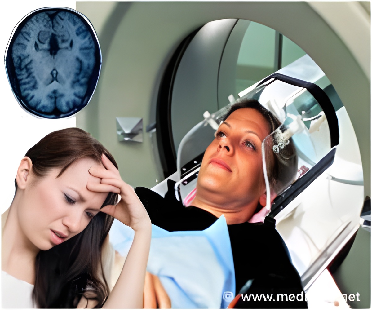
"Awareness of medical radiation exposure has increased over the past several years. In particular, the concern of additional radiation dose associated with CT in PET/CT is magnified in scans performed for investigational purposes," says Jae Sung Lee, Ph.D., associate professor of nuclear medicine at Seoul National University, Seoul, Korea.
Radiation is a natural phenomenon that is happening all the time as certain materials decay. Every year people are exposed to about 3 mSv (a measurement of exposure to radiation) of natural background radiation that comes from soil, from rocks or from high altitudes, to name some examples. As a comparison, people receive about 2 mSv during an average CT scan of the head.
"Computed tomography is useful for reducing PET scan time and improving image quality, but there is room for reducing the radiation dose—especially in brain PET studies—by avoiding the redundancy of repetitive CT scans," says Lee. "In this study, we propose a scheme to minimize the radiation dose by performing only a single CT scan per each subject and employing an image registration technique between brain PET scans."
During the study, researchers worked with five volunteers who were imaged in two sessions of dynamic brain PET/CT scanning with a new biomarker for amyloid plaque, which is implicated in cognitive decline and Alzheimer's disease. After an intermission during imaging, subjects were placed in a different position and another imaging study was performed. Both studies were 80-90 minutes long. Scientists compared images from the second PET study using the original CT image, the realigned CT image and the second CT to determine if using only the first CT scan was feasible. Researchers concluded that one CT image could be used during multiple PET studies to achieve satisfactory image quality.
Source-Eurekalert


