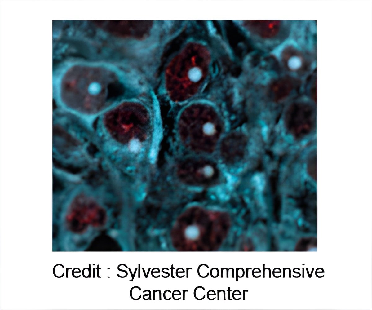
‘Anti-cancer drugs that bind to a membrane protein called dehydroorotate dehydrogenase (DHODH) can kill cancer cells.’
Tweet it Now
The Erik Marklund group at the Department of Chemistry, Uppsala University, has together with the groups of Sir David Lane and Sonia Lain at Karolinska Institutet used computer simulations together with native mass spectrometry, a technique where a protein is gently removed from its normal environment and accelerated into a vacuum chamber. By measuring the time it takes for the protein to fly through the chamber, it is possible to determine its exact weight. The research team used this highly accurate 'molecular scale' to see how lipids (the building blocks of the cell membrane) and drugs bind to DHODH."To our surprise, we saw that one drug seemed to bind better to the enzyme when lipid-like molecules were present," says assistant professor Michael Landreh, Karolinska Institutet.
The team also found that DHODH binds a particular kind of lipid present in the cell's power plant, the mitochondrial respiratory chain complex.
"This means the enzyme might use special lipids to find its correct place on the membrane," Michael Landreh explains.
To understand why lipids can help a drug recognize its target, Erik Marklund and Joana Costeira-Paulo employed computer simulations to explore the structure and dynamics of free as well as membrane-bound DHODH.
Advertisement
"The study helps to explain why some drugs bind differently to isolated proteins and proteins that are inside cells. By studying the native structures and mechanisms for cancer targets, it may become possible to exploit their most distinct features to design new, more selective therapeutics," says Sir David Lane, Karolinska Institutet.
Advertisement
Source-Eurekalert


