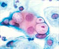
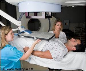
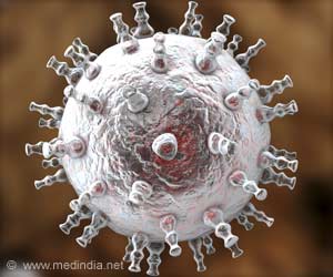
The pathology using infrared spectroscopic imaging doesn’t require any actual staining to analyze the chemical composition of cells.
The technique uses infrared spectroscopic imaging to analyze the chemical composition of cells directly. The investigators were able to identify correlations that a computer can use to do quick pathology analysis by noting the spectral characteristics of light bouncing off the cells.
The scientists hope that the new method will drastically increase the speed of digital pathology studies, since manual aspects of staining and preparation may be avoided altogether. The technology relies on computation, instead of staining to provide images.
"The imaging allows histology digital imaging without destroying tissue properties caused by staining, therefore the same slide can be used for other purposes. For research applications, it also allows higher throughput by rapidly marking up the tissues for regions of interest,” said David Mayerich, the lead author of the study.
Source-Medindia

