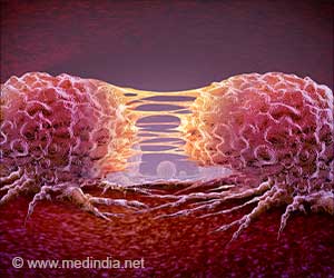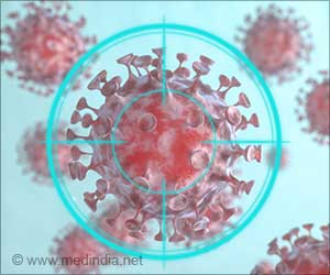
‘Nanotag, the newly developed nanoparticle produces 10 times greater signal enhancement making it possible to detect minute amounts of organic molecules, such as DNA, for particular diseases.’
Tweet it Now
"It was calculations that no one on campus had done before," explained Sagle, who serves as an advisor to Gorunmez. "Zohre, essentially by herself and without a lot of guidance and help, got these calculations up and running." The discovery came in 2013 as part of the Sagle Lab research group's work in developing new methods to study and examine single molecules using a technique called surface-enhanced Raman spectroscopy, or SERS.
The technique targets molecules using lasers, which results in a scattering of light at different wavelengths along a spectrum. Because the molecules produce weak signals, gold or silver nanoparticles are used to amplify them, which is measured by a spectrometer for analysis.
The process is highly sensitive and fraught with challenges, including difficulties with reproducibility, signal stability and a lack of quantitative information.
The team looked to previous research, which showed greater enhancement from molecules residing within a one nanometer gap between a structure with a smooth metallic core and shell. But this one nanometer gap - 100,000 times smaller than the width of a human hair - is often difficult and expensive to produce, resulting in a lack of widespread use.
Advertisement
The newly created nanotag produced 10 times greater signal enhancement compared to smooth-shell core structures, making it possible to detect minute amounts of organic molecules, such as DNA, for particular diseases, she said.
Advertisement
"This allows you to target, image and release drugs all with one device," she explained.
While the discovery itself proved novel, Sagle knew that the team's promising nanotag needed to be further analyzed, understood and modeled before it could be used in biological applications.
Source-Eurekalert


