A novel image-processing algorithm makes use of 20-times less radiation to produce the information needed by radiologists.
Perfusion CT scanning, an emerging imaging technology, was slammed in 2009 when a machine set to incorrect radiation levels overdosed hundreds of people in Los Angeles.
Advertisement
After the incident, researchers at the Mayo Clinic have developed a way to reduce the amount of radiation involved in the procedure which, when done properly, already involves very little risk.
At the correct dose, there should be no injury," said Cynthia McCollough.
"We believe in the clinical value of perfusion CT, so we're trying to lower the dose and reduce the stigma," she added.
McCollough and her colleagues created a new image-processing algorithm that can give radiologists all of the information they need using as up to 20 times less radiation, depending on the diagnostic application.
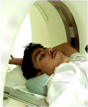
A typical CT perfusion procedure lasts about half a minute and scans the same tissue many times, each scan at a low dose.
"When we use very low doses, the noise gets so high that it's hard to tell what you are seeing," said Juan Carlos Ramirez Giraldo.
"With this algorithm, we're trying to maintain both the image quality, so that a doctor can recognize the anatomic structures, and the functional information, which is conveyed by analyzing the flow of the contrast agent over the many low dose scans," he added.
"We're up to 15 or 20 cases that we've shown to the docs, and they're all giving us the thumbs up," said McCollough.
The research will be presented at the 52nd Annual Meeting of the American Association of Physicists in Medicine (AAPM) in Philadelphia.
Source: ANI
"We believe in the clinical value of perfusion CT, so we're trying to lower the dose and reduce the stigma," she added.
Advertisement
McCollough and her colleagues created a new image-processing algorithm that can give radiologists all of the information they need using as up to 20 times less radiation, depending on the diagnostic application.
A typical CT perfusion procedure lasts about half a minute and scans the same tissue many times, each scan at a low dose.
"When we use very low doses, the noise gets so high that it's hard to tell what you are seeing," said Juan Carlos Ramirez Giraldo.
"With this algorithm, we're trying to maintain both the image quality, so that a doctor can recognize the anatomic structures, and the functional information, which is conveyed by analyzing the flow of the contrast agent over the many low dose scans," he added.
"We're up to 15 or 20 cases that we've shown to the docs, and they're all giving us the thumbs up," said McCollough.
The research will be presented at the 52nd Annual Meeting of the American Association of Physicists in Medicine (AAPM) in Philadelphia.
Source: ANI
Advertisement
Advertisement
|
Advertisement
Recommended Reading
Latest Research News

The scientists discovered that when the stem cells in the hair follicle are made softer, they have a higher chance of growing hair.

A combinatorial therapy provided promising beneficial results among people with ischemic stroke.

In Parkinson's disease patients voice disorders are quite common. A new combination therapy had greater effects on the voice.

New study findings delineate how aging affects the immune response following Spinal cord injury (SCI) and highlight the participation of the spinal cord meninges in repair.

A recent clinical trial suggests that the first medication therapy to reduce the progression of nearsightedness in children could be on the way.