Highlights:
- Cancer chemotherapy causes several side effects due to its action on normal tissues of the body
- Focused ultrasound can direct nanoparticle-stabilized microbubbles containing chemotherapy to the target cancer site
- A study in triple negative breast cancer in mice has produced positive results
Since nanoparticles are too big to cross normal blood vessels, they remain in the blood stream and do not diffuse into normal tissues. The blood vessels supplying the cancer cells are more porous. The nanoparticles diffuse through these blood vessels and selectively affect cancer cells. Thus, the nanoparticles are mainly effective against the cancer cells that are close to the blood vessels.
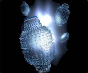
To circumvent this limitation and enhance deeper penetration of the nanoparticles into cancerous tissues, scientists came up with the approach of using focused ultrasound and nanoparticle-stabilized microbubbles. They conducted their study on mice implanted with triple negative breast cancer tissue. Triple negative breast cancer is resistant to several treatments which include the estrogen and progesterone receptor antagonists and the HER 2 receptor antagonists. For the experiment, the twelve mice with the cancer were divided into three groups:
- The first group did not receive any treatment and served as a control. In this group, the cancer continued to grow, as expected.
- The second group was treated with the nanoparticle-stabilized microbubbles that contained the anticancer drug cabazitaxel. Cabazitaxel is currently approved by the US Food and Drug Administration for the treatment of metastatic prostate cancer that has not responded to hormone therapy. The scientists had established that the drug is effective against the cancer cell lines used in the study. The animals in this group responded, albeit variably, to the treatment. The tumors started re-growing 50 days after the treatment was stopped.
- The third group was treated with cabazitaxel-containing nanoparticle-stabilized microbubbles. This was followed by the application of ultrasound with a high acoustic pressure (mechanical index of 0.5). The ultrasound caused the bubbles to vibrate, burst and release the nanoparticles. It also made the blood vessels more porous, thus promoting the passage of the nanoparticles deeper into the cancer tissue, which was confirmed using imaging studies. The acoustic pressure (MI=0.5) of the ultrasound that was used was tolerable; lower pressures were ineffective and very high pressures (mechanical index of 1) could cause bleeds in the eye.
Focused ultrasound is non-invasive and painless. Due to the selective action on the tumor, a larger amount of the chemotherapy drug can be administered without causing toxic effects. This technique could also have possible benefits in brain cancers, where it could increase the permeability of the blood-brain barrier and enable the anticancer drugs to reach the brain cancer.
The use of focused ultrasound and microbubbles to deliver the chemotherapy drug gemcitabine for the treatment of locally advanced pancreatic cancer is being tried out in human clinical trials. Thus, it may not be too long before such treatments become a reality.
- Snipstad S et al. Ultrasound Improves the Delivery and Therapeutic Effect of Nanoparticle-Stabilized Microbubbles in Breast Cancer Xenografts. Ultrasound in Medicine and Biology. DOI: http://dx.doi.org/10.1016/j.ultrasmedbio.2017.06.029
Source-Medindia


