‘μChIP-seq method helps investigate how epigenetic information is passed from one generation to the next to orchestrate the MZT (maternal to zygote transition).’
Tweet it Now
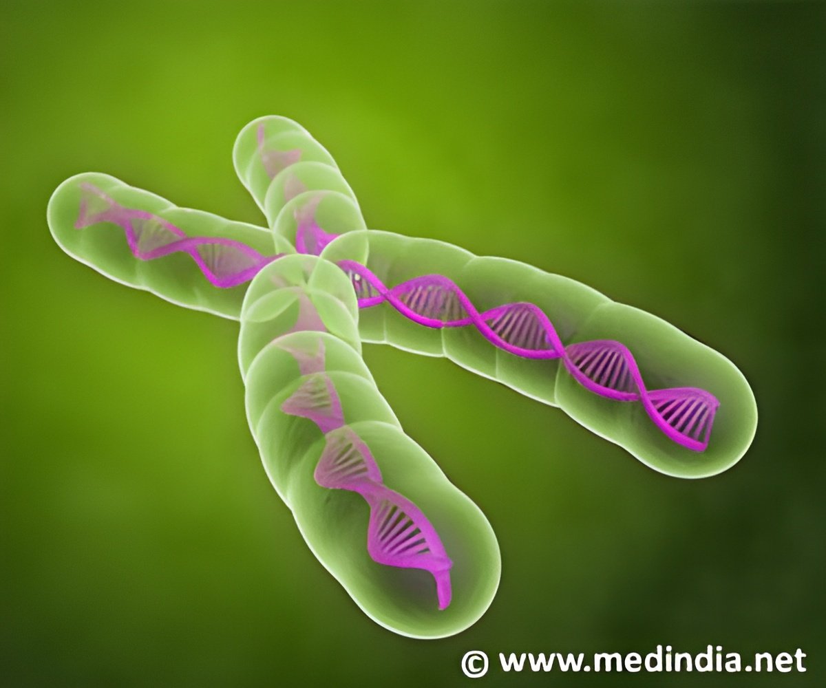
The improved sensitivity of μChIP-seq, developed jointly by Klugland, Ren and John Arne Dahl of Oslo University, led to “some remarkable discoveries that have completely changed our view of epigenetic inheritance mechanisms,” says Ren.In the current paper, Ren and his colleagues describe their use of μChIP-seq to investigate how epigenetic information is passed from one generation to the next to orchestrate the MZT. To that end, they examined in mouse embryos the global distribution of an epigenetic mark known to play a critical role in regulating the activity of genes. This mark (H3K4me3) modifies chromatin—the complex of DNA and its protein packaging—by adding three identical molecules known as methyl groups at a specific place on a packaging protein known as histone 3.
Dahl, a visiting scholar in Ren’s lab and co-first author on the Nature paper, fine-tuned the technique for mapping such epigenetic tags to where only a few hundred embryos or cells were needed for each experiment. Previous methods required up to 10,000 cells to conduct similar analyses. The other first author, Inkyung Jung, a Ludwig postdoc in Ren’s lab, contributed significantly to the computational analysis of the resulting data.
When the scientists compared H3K4me3 distribution in immature mouse egg cells they found something unexpected: broad but distinct domains of the immature egg cell’s genome, representing some 22% of the whole, are heavily marked by H3K4me3. These domains rapidly decrease in size in 2-cell embryos and eventually shrink to about 1% to 2% of the genome.
“The key lesson we learned was that the genes that are destined to be turned on specifically in the fertilized egg are covered by this unique chromatin domain structure,” says Ren. Those marks have to be removed by specialized enzymes to activate the zygotic genome. “This mark is a mechanism for the oocyte to influence which genes in the zygote are activated. It is an epigenetic mechanism for the passage of information from the maternal oocyte to the zygote.”
Advertisement
“With a better understanding of the epigenetic landscapes in cancers, we are going to have more tools to study the basis of tumorigenesis,” says Ren. “We still have a long way to go, but our goal is to have a thorough understanding of gene regulatory programs so we can use that knowledge to treat cancer and develop diagnostic tools.”
Advertisement
Bing Ren is a member of the Ludwig Institute for Cancer Research, San Diego, and a professor of cellular and molecular medicine at the University of California, San Diego, School of Medicine.
Source-Newswise

