A device that will enable early detection of eye disease in premature babies has been developed by researchers at Duke University Medical Center.
The technology uses spectral domain optical coherence tomography (SD OCT) to create a 3-D picture of the back of the eye."This new tool is changing the way we identify eye conditions in infants," says Cynthia Toth, MD, an ophthalmologist at the Duke Eye Center, who is leading the study that appears online this month in the journal Ophthalmology.
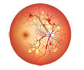
Retinopathy of prematurity (ROP) is one of the most common causes of vision loss in children. It occurs when babies are born prematurely and their retinal blood vessels don't develop fully. Instead, the vessels grow abnormally and are prone to leaking and contracting. That can pull the retina out of position, causing retinal detachments that can lead to visual loss and blindness.
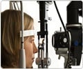
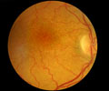
Current screening for ROP is based on two-dimensional images taken either with an ophthalmoscope or a camera placed directly on the infant's cornea.
"Examining the retina with these methods is like looking at the surface of the ocean and only seeing dimly into the shallow water," says Toth, a professor of ophthalmology and biomedical engineering.
"You cannot see what lies below," the expert added.
Advertisement
"This is like looking into an aquarium from the side, where all the fish at every depth are visible," Toth says.
Advertisement
TAN


