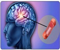
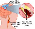
Susceptibility-weighted imaging (SWI) is a powerful tool for characterizing infarctions (stroke) in patients earlier and directing more prompt treatment, says a new study.
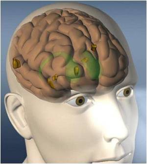
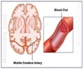
For the study, researchers assessed that of the 35 patients with thromboembolic infarctions, SWI detected thromboemboli in 30 patients. Additionally, 14 of these thromboemboli were located in arteries other than the anterior division of the MCA. Dr. Mamlouk said, "At our institution, we are amazed at how often SWI detects thromboemboli in all major cerebral arteries, not just the MCA. Given SWI's high sensitivity (86%) of thromboemboli detection, we found that there is an adjunctive role of SWI in classifying cerebral infarctions in patients."
While MRIs have been the gold standard for evaluating infarctions, adding SWI to the routine MRI sequences for evaluating patients with a clinical suspicion of stroke will hasten their time to treatment and improve overall recovery, said Dr. Anton Hasso, senior author of the study. Dr. Mamlouk states, "The utility of SWI extends beyond the evaluation of hemorrhage. Using SWI in patients with cerebral infarctions will decrease further imaging and its associated costs and radiation exposure, but more importantly this imaging technique will guide direct management in a timelier manner."
Dr. Mamlouk will deliver a presentation on this study on Wednesday, May 4, 2011 at the 2011 ARRS Annual Meeting at the Hyatt Regency Chicago.
Source-Eurekalert

