‘New computer modeling may help doctors and eye specialists to learn more about the risk factors and causes of glaucoma, which is a progressive disease that causes damage to the optic nerve in the eye.’
Read More..Tweet it Now
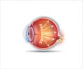
Glaucoma is a progressive disease that causes damage to the optic nerve in the eye. Abnormally high pressure of fluids in the eye (intraocular pressure or IOP) may lead to glaucoma and can cause irreversible vision loss if not treated. However, clinicians may not always know the underlying cause of increases in IOP or understand how changes in IOP affect the rate of blood flow (blood velocity) in the eye.Read More..
Using data from scientific literature on IOP and measurements of the diameter, thickness and elasticity of the vessel walls inside the eye, researchers developed a reduced-dimension model of ocular circulation to simulate blood velocity and vessel deformation. Reduced-dimension models use geometric shapes, lines and points to represent blood vessels and other physiological structures. This kind of model captures the main mechanisms of body systems while solving the given problem easily and quickly on a computer, providing results that could be integrated in a clinical setting.
This model can be used as a “virtual toolbox” to help eye clinicians learn more about the mechanisms and factors that contribute to the development and progression of conditions such as glaucoma, explained Lucia Carichino, PhD, of the Rochester Institute of Technology in New York. Carichino’s model indicates that the tiny blood vessels in the eye (venules) get smaller as IOP increases. The venules’ reduced diameter then affects circulation in the central retinal artery, a larger vessel that provides nutrients to the eye and is often used to measure ocular blood flow.
“Mathematical tools can be used in synergy with clinical data to significantly advance the understanding of the mechanisms that affect the blood flow in the eye,” Carichino said. The use of this model “suggests that the retinal microvasculature, in particular retinal venules, plays a fundamental role in studying ocular blood flow,” she added.
Source-Newswise


