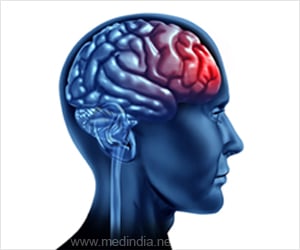
Brains of adults suffering from autism are wired differently than their normal counterparts and could explain the social issues faced by the former, researchers have said.
Scientists at the University of Washington's Autism Center used functional magnetic resonance imaging to discover that the most severely socially impaired subjects in the study displayed the most abnormal pattern of connectivity among a network of brain regions involved in face processing."This study shows that these brain regions are failing to work together efficiently. Our work seems to indicate that the brain pathways of people with autism are not completely disconnected, but they are not as strong as in people without autism," said Natalia Kleinhans, a research assistant professor of radiology and lead author of the paper published in the journal Brain.
This is the first study to deal with brain connectivity and social impairment, focussing on how the brain processes information about faces. Facial discrepancies are one of the earliest characteristics to emerge in people with autism.
Led by Elizabeth Aylward, a UW professor of radiology, the research team examined connectivity in the limbic system, or the network of brain regions that are involved with processing social and emotional information. The study was conducted on 19 high-functioning adults with autism in the age group of 18 to 44 having IQs of at least 85. They were then compared with an age- and intelligence-matched sample of 21 typically developed adults.
The group with autism spectrum disorder included 8 individuals diagnosed with autism, nine with Asperger's syndrome and two diagnosed with pervasive developmental disorder not otherwise specified. The level of social impairment for each autistic participant was taken from records of clinical observations and diagnoses that confirmed that each had autism.
The researchers scanned brains of each participant while looking at pictures of faces or houses. Participants were shown four series of 12 pictures of faces and a similar number of series showing houses. Each picture was seen for three seconds. Occasionally the same face or house picture was repeated, and participants were told to press a button when this occurred.
Advertisement
The researchers focused on the fusiform face area of the brain, a region that is involved in face identification. Compared to the participants with autism, the typically developing adults showed significantly more connectivity between the fusiform face area and two other brain regions, the left amygdala and the posterior cingulate.
Advertisement
"This study shows that the brains of people with autism are not working as cohesively as those of people without autism when they are looking at faces and processing information about them," said Kleinhans.
Source-ANI
RAS/L


