Medindia LOGIN REGISTER
Medindia
Advertisement

Brain Abnormalities in Migraine Patients

by Sheela Philomena on March 26, 2013 at 12:16 PM
 Brain Abnormalities in Migraine Patients

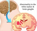
Magnetic resonance imaging shows the presence of brain abnormalities that are related to migraines. The research is published online in the journal Radiology.

Migraines are intense, throbbing headaches, sometimes accompanied by nausea, vomiting and sensitivity to light. Some patients experience auras, a change in visual or sensory function that precedes or occurs during the migraine. More than 300 million people suffer from migraines worldwide, according to the World Health Organization.

Advertisement

Previous research on migraine patients has shown atrophy of cortical regions in the brain related to pain processing, possibly due to chronic stimulation of those areas. Cortical refers to the cortex, or outer layer of the brain.

Much of that research has relied on voxel-based morphometry, which provides estimates of the brain's cortical volume. In the new study, Italian researchers used a different approach: a surface-based MRI method to measure cortical thickness.
Advertisement

"For the first time, we assessed cortical thickness and surface area abnormalities in patients with migraine, which are two components of cortical volume that provide different and complementary pieces of information," said Massimo Filippi, M.D., director of the Neuroimaging Research Unit at the University Ospedale San Raffaele and professor of neurology at the University Vita-Salute's San Raffaele Scientific Institute in Milan. "Indeed, cortical surface area increases dramatically during late fetal development as a consequence of cortical folding, while cortical thickness changes dynamically throughout the entire life span as a consequence of development and disease."

Dr. Filippi and colleagues used magnetic resonance imaging (MRI) to acquire T2-weighted and 3-D T1-weighted brain images from 63 migraine patients and 18 healthy controls. Using special software and statistical analysis, they estimated cortical thickness and surface area and correlated it with the patients' clinical and radiologic characteristics.

Compared to controls, migraine patients showed reduced cortical thickness and surface area in regions related to pain processing. There was only minimal anatomical overlap of cortical thickness and cortical surface area abnormalities, with cortical surface area abnormalities being more pronounced and distributed than cortical thickness abnormalities. The presence of aura and white matter hyperintensities—areas of high intensity on MRI that appear to be more common in people with migraine—was related to the regional distribution of cortical thickness and surface area abnormalities, but not to disease duration and attack frequency.

"The most important finding of our study was that cortical abnormalities that occur in patients with migraine are a result of the balance between an intrinsic predisposition, as suggested by cortical surface area modification, and disease-related processes, as indicated by cortical thickness abnormalities," Dr. Filippi said. "Accurate measurements of cortical abnormalities could help characterize migraine patients better and improve understanding of the pathophysiological processes underlying the condition."

Additional research is needed to fully understand the meaning of cortical abnormalities in the pain processing areas of migraine patients, according to Dr. Filippi.

"Whether the abnormalities are a consequence of the repetition of migraine attacks or represent an anatomical signature that predisposes to the development of the disease is still debated," he said. "In my opinion, they might contribute to make migraine patients more susceptible to pain and to an abnormal processing of painful conditions and stimuli."

The researchers are conducting a longitudinal study of the patient group to see if their cortical abnormalities are stable or tend to worsen over the course of the disease. They are also studying the effects of treatments on the observed modifications of cortical folding and looking at pediatric patients with migraine to assess whether the abnormalities represent a biomarker of the disease.

Source: Eurekalert
Font : A-A+

Advertisement

Advertisement
Advertisement

Recommended Readings

Latest Research News

Can Sleep Brain Waves Defend Against Epileptic Activity?
Memory deficits in individuals with epilepsy, especially cognitive difficulties, might partially stem from the transient impairments caused by these slow waves.
High Altitude Training's Impact on Surgical Readiness
High altitude training exhibits potential benefits for pre-surgery patients, enhancing readiness, stated a new study.
AI Shows Promise in Identifying ADHD
The AI method shows promise in identifying imaging biomarkers for diagnosing ADHD.
Life Expectancy Gap for Autistic Individuals Revealed
Diagnosed autistic individuals showed increased premature mortality in the UK, highlighting urgent needs to address associated inequalities.
Exploring How Hearing Impairment Shapes Dementia Risk
Study reveals a correlation between hearing impairment and distinct brain region variances, contributing to dementia.
View All
This site uses cookies to deliver our services.By using our site, you acknowledge that you have read and understand our Cookie Policy, Privacy Policy, and our Terms of Use  Ok, Got it. Close
×

Brain Abnormalities in Migraine Patients Personalised Printable Document (PDF)

Please complete this form and we'll send you a personalised information that is requested

You may use this for your own reference or forward it to your friends.

Please use the information prudently. If you are not a medical doctor please remember to consult your healthcare provider as this information is not a substitute for professional advice.

Name *

Email Address *

Country *

Areas of Interests