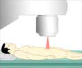
Lipids, water, oxyhemoglobin and deoxyhemoglobin in the blood all respond to laser light, said Dr. Dogra. "By observing increases and decreases in these four things, we can tell if the tissue is malignant or benign, he said. Deoxyhemoglobin is the biggest distinguisher between malignant and benign. If deoxyhemoglobin increases even slightly in intensity, the odds that the tissue is malignant increases dramatically," he said.
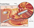
Prostate cancer is the second leading cause of cancer death in American men. Transrectal ultrasound, the current gold standard to diagnose prostate cancer, has an overall success rate of about 70%, said Dr. Dogra. "Transrectal ultrasound is an invasive procedure and most men do not like it. There is a need for a new imaging technique," Dr. Dogra said. "We expect this technique to be clinically available in about five years," he added.
Dr. Dogra will present his study at the ARRS annual meeting on April 18 in Washington, DC.
Source-Eurekalert


![Prostate Specific Antigen [PSA] & Prostate Cancer Diagnosis Prostate Specific Antigen [PSA] & Prostate Cancer Diagnosis](https://www.medindia.net/images/common/patientinfo/120_100/prostate-specific-antigen.jpg)


