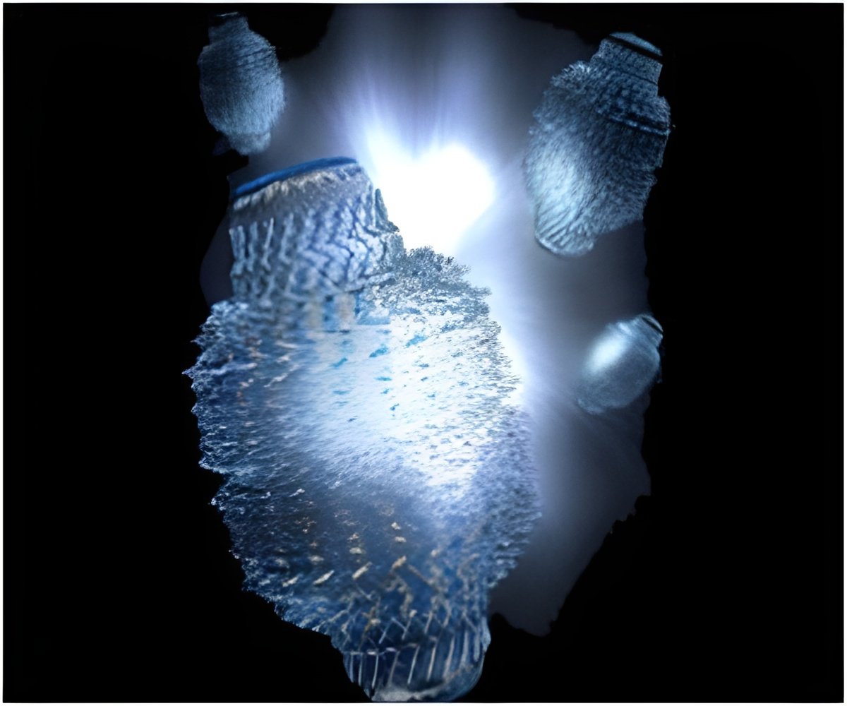
A University of Strathclyde academic has been harnessed by a pioneering imaging technique to track the effects of next-generation nanomedicines on patients.

Professor Kumar, whose team's research article has been published in the journal PLOS ONE, said: "Nanotechnology's role in drug delivery has the power to transform the way patients are given medicines over the next decade or so."
"In the case of traditional medicines, such as tablets and capsules, only a limited amount of drug – thought to be around five to 15 per cent for the majority of compounds – makes it through the gut into patients' blood." The good thing about nanomedicines is that – unlike as is the case with traditional tablets and capsules – the drugs are not released in the gut. Instead, nanomedicines are absorbed intact and release the encapsulated drugs directly into bodily tissues, including the blood, offering the possibility to reduce the required dose without compromising the therapeutic effects.
"All medicines are combined with what are known as 'excipients' – inactive substances which give them the desired bulk and consistency and their role is restricted to the gut." However, the excipients such as polymers, used to formulate the nanoparticle-encapsulating drugs may exhibit undesired effects when they are absorbed through the gut wall. Scientists want to know if nanoparticle-based drugs can have any adverse effects on patients – and, in particular, if they cause more harm than good in some cases.
"Up until now, little has been known about what happens after nanoparticles circulate throughout the body and if they raise any safety issues for the patient. Previously, it was necessary for nanoparticles to be given a fluorescent or radioactive label, in order to allow scientists to be able to identify and track them. However, by using PeakForce QNM atomic force microscopy we can, for the first time, track where these nanoparticles are going throughout the body after oral administration – without attaching any fluorescent or radioactive labels and by using the real drug loaded nanoparticles. In particular, we can identify if they are accumulating in specific areas, causing what is known as 'tissue stiffness' – a condition linked to a variety of diseases, including cancer."
Professor Kumar said it is known that tumours are more rigid – or stiff – when compared with surrounding healthy tissues. In addition, recent studies using atomic force microscopy have also shown it is possible to distinguish between non-malignant and malignant tumours cells, on the basis of their relative stiffness.
Advertisement
"By using atomic force microscopy in this way, we may in future be able to analyse patients' blood and tell if, for example, nanomaterials are accumulating in their livers or arterial walls, causing stiffness which – if it persists long enough – may increase their chances of developing diseases."
Advertisement
Source-Eurekalert


