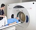
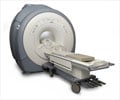
American researchers have for the first time combined two kinds of body imaging in a single scanner, i.e. positron emission tomography (PET) and magnetic resonance imaging (MRI).
Simon Cherry, a professor at the University of California, Davis who led the project, said that the revolutionary scanner was built for studies with laboratory mice, for example in cancer research.Highlighting the importance of the new scanner, he said that MRI scans could provide exquisite structural detail but little functional information, while PET scans could show body processes but not structures.
"We can correlate the structure of a tumour by MRI with the functional information from PET, and understand what's happening inside a tumour," Cherry said.
Working out a combination of the two types of scan in a single machine is difficult because they interfere with each other. While the magnetic fields on which MRI scanners rely can easily be disturbed by metallic objects inside the scanner, they may seriously effect the detectors and electronics needed for PET scanning.
According to Cherry, there is also a limited amount of space within the scanner in which to fit everything together.
The researchers used a new technology, the silicon avalanche photodiode detector, in their machine.
Advertisement
A paper describing the new scanner ha sbeen published online in the journal Proceedings of the National Academy of Sciences.
Source-ANI
SRM /J


