- GHAI essential pediatrics - 8th edition
- Harrison's principles of internal medicine -volume 2- 18th edition
- Transposition of the great arteries - (http://www.nlm.nih.gov/medlineplus/ency/article/001568.htm)
What is Transposition of Great Vessels?
Transposition of great vessels / transposition of great arteries (TGA) is a condition where the great vessels (aorta & pulmonary artery) arise from inappropriate ventricles of the heart.
It is a congenital heart anomaly, that is, the structural heart defect is present at birth. It is also classified as a cyanotic heart disease since it causes bluish discoloration of skin, nails, mucous membranes etc. due to reduced oxygen content of the blood.
What is the Problem in Transposition of the Great Arteries?
To understand the pathology of transposition of great arteries, we must first understand the normal structure and functioning of human heart. The human heart is a four-chambered muscular structure about the size of the fist and it pumps blood. The upper two chambers are called atria (right atrium & left atrium), which are separated from each other by a membrane called as inter-atrial septum. The lower two chambers are called ventricles (right & left ventricle), which are separated from each other by the interventricular septum. Normally, the right ventricle gives rise to the pulmonary artery. The pulmonary artery carries away the deoxygenated blood from the right ventricle of heart to the lungs and gets it oxygenated. This is called pulmonary circulation. Also normally, the left ventricle gives rise to the aorta. The aorta carries away the oxygenated blood from the left ventricle of heart and distributes it to the entire body. This is called systemic circulation. The aorta and the pulmonary artery are referred to as the great arteries.
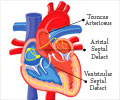
Transposition of Great Arteries
Transposition of great arteries is a congenital heart disease in which, the great vessels arise from inappropriate ventricles. That is:
- Aorta arises from the right ventricle
- Pulmonary arteries arise from the left ventricle
In the fetal heart, there is opening in the inter-atrial septum called as foramen ovale, which allows mixing of blood during fetal life. After birth, the lungs expand and the baby starts to breath. The blood now gets oxygenated by lungs and there is no need for the foramen ovale to remain open. So, after birth, the foramen ovale closes immediately. If it remains open, it results in a defect in the atrial septum called atrial septal defect or patent foramen ovale. A similar defect can also occur in the ventricular septum, referred to as ventricular septal defect or VSD. The defects in the atrial or ventricular septa are commonly referred to as a ‘hole in the heart’. Some babies may have a communication between the aorta and the pulmonary artery after birth, called patent ductus arteriosus.
In transposition of great vessels, blood from the heart enters the lungs and gets oxygenated, then it returns to the heart and then again to the lungs. The cycle continues and the blood in this cycle does not go to the rest of the body. In another cycle of blood circulation (systemic), blood from the body reaches heart and again gets distributed in the body without getting oxygenated in lungs. If the baby has an ASD or VSD, there is a good chance that some mixing of blood will take place between oxygenated and deoxygenated blood in the heart. An ASD is usually small and the mixing is poor. Presence of VSD of adequate size results in good mixing, but still life cannot be maintained for a longer time and heart failure occurs in cases of TGA with VSD at around 4-10 weeks after birth.
What are the Causes of Transposition of Great Vessels?
Conditions that increase the risk of a baby developing transposition of great vessels include the following:
- Insulin dependent maternal diabetes during pregnancy
- Maternal age over 40 yrs
- Poor nutrition during pregnancy
- Ingestion of certain drugs such as benzodiazepines during pregnancy
- Infection with rubella (German measles) or other virus during pregnancy
What are the Symptoms & Signs of Transposition of Great Vessels?
Symptoms and signs noted in a baby with transposition of great vessels are as follows:
- Babies with complete TGA with intact interventricular septum are blue at birth. Babies with a VSD may become blue a few weeks after birth. The bluish discoloration is referred to as cyanosis.
- Rapid breathing
- Lack of appetite and poor weight gain
- In TGA without VSD, heart failure develops within the first week of life but in patients of TGA with VSD, heart failure develops around 4-10 weeks of life.
- The heart size can be normal in the first two weeks of life but enlarges rapidly
How to Diagnose Transposition of Great Vessels?
Transposition of great vessels is diagnosed based on:
- General examination, which may reveal
- Cyanosis
- Rapid breathing
- Auscultation, or examination of the heart with a stethoscope. The doctor may be able to appreciate certain sounds which indicate heart disease
- First heart sound- normal
- Single second heart sound
- Grade 1-2 ejection systolic murmur in TGA without VSD
- Grade 2-4 ejection systolic murmur in TGA with VSD
- Chest x-rays, which reveal
- Cardiomegaly or enlargement of the heart
- Plethoric lung fields, or prominent blood vessels in the lungs
- Cardiac MRI
How is Transposition of Great Vessels Treated?
Unfortunately, TGA is a fatal disorder if left untreated and the only treatment is surgical intervention by highly specialized pediatric cardiac surgeons.
- Prostaglandin E1 can help reduce the cyanosis, by keeping atrial communication open.
- Atrial communication can also be kept open by a procedure called “balloon atrial septostomy”. Septostomy is successful only up to the age of 6-12 weeks and gives temporary relief by providing better mixing and reducing left atrial pressure.
- “Arterial switch operation” is now established as the treatment of choice for TGA.
- The pulmonary artery & aorta are cut off
- Then they are attached at appropriate places. That is, the positions of misplaced arteries are switched to their correct places
- The time period or window for getting this surgery done is very crucial. Infants of TGA without VSD should get this surgery done within the first 2-4 weeks of life
- Twenty-year survival is >90 %
- “Atrial switch operation / Senning operation” – Later in infancy, this is the only option of surgery for patients of TGA without VSD. But, this is not an ideal long-term operation.


