Diagnosis
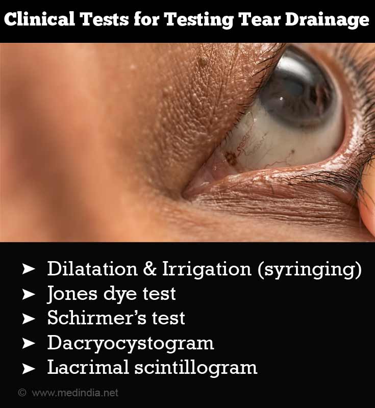
Clinical tests for testing tear drainage are as follows: Dilatation & Irrigation (syringing), Johne’s dye test, Schirmer’s test, dacryocystogram and lacrimal scintillogram.
The eye doctor will ask questions to find out any evidence of eye, sinus or dental disease which may be causing watery eyes.
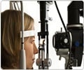
He then does a detailed examination of the eye under magnification of slit lamp to observe for blepharitis, conjunctivitis, keratitis or a foreign body lodged beneath the lids.
The position of the lids and the tear drainage opening is carefully observed.
The clinician then performs the following tests -
Dilatation of the punctum (tear drainage opening) - This is done in cases of stenosed or narrowed punctum. The test helps to diagnose as well as to treat the condition. It is done with a sleek instrument with a pointed tip gradually increasing in girth. As it passes down the narrowed punctum, it dilates the punctum and restores the drainage.
Syringing - A fine cannula is inserted in each of the puncta and water is irrigated through thecanula. This is done to check the patency of the tear drainage system of the eye. If the water enters the patient’s nose or the back of the throat, the passageway is considered patent.
If the water does not enter the throat and simply flows out through the opposite punctum, it suggests that the watery eye is due to blocked tear drainage pathway.
Johne’s Dye test - This test is performed by inserting a small amount of fluorescent dye in the eye and noting whether it drains in the nose. It is helpful in determining and differentiating between the drainage pump failure and partial block. Differentiating between these conditions helps to guide treatment.
Lacrimal scintilography - It is a test done by instilling a drop of radioactive Technitium 99 into the eye and tracing its path. This test helps to detect functional and anatomical site of obstruction.
- On the left side the tracer was seen to enter the nose.
- On the right side tracer not seen in the nose with majority of the tracer being held up proximally. Indicating an obstruction.
Dacryocystography - It is done by injecting a contrast dye into the drainage cannaliculi and observing its path radiologically. This test helps to pinpoint the site of blockage in tear drainage pathway.
- Bilateral dacryocystography (DCG) with complete obstruction of the left canaliculus.
Schirmer’s test - It is done by placing a filter paper 5mm X 35mm sized into the eye and noting its wetness after 5 mins. This test helps to diagnose dry eye and its severity from the amount of wetting of the filter paper.
- Schirmer’s test with strips placed in situ


