- Spinal Stenosis - (https://medlineplus.gov/spinalstenosis.html)
- Cauda equina syndrome - (https://en.wikipedia.org/wiki/Cauda_equina_syndrome)
What is Spinal Stenosis?
Spinal stenosis refers to narrowing of the vertebral or spinal canal which encloses the spinal cord and spinal nerves. It may result in compression of these nerves leading to pain, numbness, tingling and muscle weakness affecting the lower back, legs or the arms and shoulder depending on where the canal stenosis has occurred.
Spinal stenosis most commonly affects the lower part of the spine (lumbar spine stenosis) or the cervical (neck). It occurs due to wear and tear of the spine with age and is mostly seen in persons over the age of 50 years. It may occur in younger individuals born with a narrow spinal canal or following trauma.
Although there is no cure, most cases respond to conservative measures such as pain medication and exercise and weight reduction. If these measures do not help, steroid injections or surgery may be necessary.
Spinal Anatomy and its Effects on Types of Spinal Stenosis
To understand about spinal stenosis, it is necessary to know in brief about the structure and function of the spinal canal. The vertebral column or spine is made up of 33 bones or vertebrae arranged one below the other. It extends from the neck above to the sacrum below.
Successive vertebrae are separated by intervertebral discs (cartilage) that act as shock absorbers and prevent the vertebrae from rubbing against one another.
Each vertebra has an anterior part called the body and a posterior arch consisting of 2 transverse processes and a spinous process at the back. In the center of each vertebra is a space called the vertebral foramen (refer figure). Vertebral foramina of all vertebrae line up to form the vertebral or spinal canal.
When viewed from the side a gap can be seen between successive vertebrae called the intervertebral foramen (nerve root canal) through which the spinal nerves emerge to branch out to various parts of the body. Both the spinal canal and the nerve root canal are surrounded by bone and connective tissue (ligaments). Bony changes and disease can cause narrowing of these canals and can compress the spinal cord or the spinal nerves as the case may be.
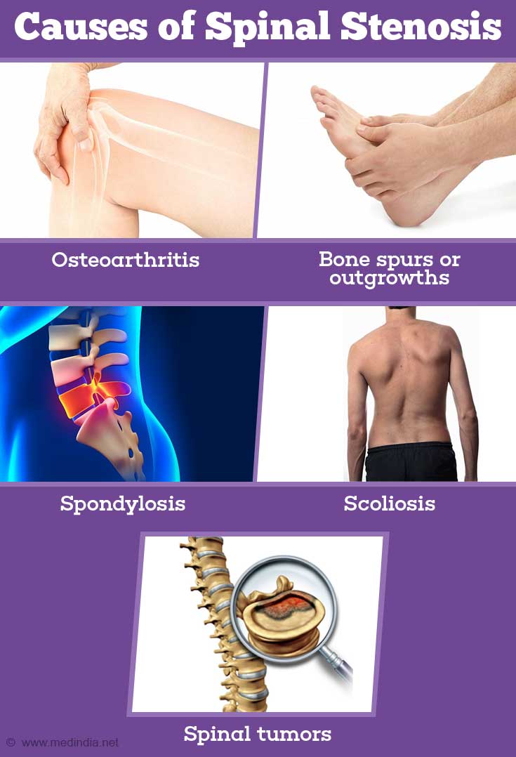
What are Causes of Spinal Stenosis?
- Degenerative changes in vertebrae due to aging such as
- Osteoarthritis (thinning of joint cartilage)
- Bulging of intervertebral disk into the canal causing narrowing or stenosis
- Bone spurs or outgrowths
- Enlargement of facet joint (synovial joint between articular processes of the vertebrae), resulting in less space available for the spinal nerves to exit the nerve root canal
- Spondylosis (abnormal fusion and immobilization of vertebral bones)
- Spondylolisthesis - Forward displacement of one of the lower lumbar vertebrae over the vertebra below it or on the sacrum
- Scoliosis (congenital or acquired) – a type of spine deformity
- Traumatic injury
- Vertebral fracture
- Dislocation
- Rheumatoid arthritis
- Paget’s disease
- Ankylosing spondylitis
- Spinal tumors
- Fluorosis (excessive levels of fluorine in the body)
- Cervical stenosis - the narrowing occurs in the cervical or neck portion of the spine
- Lumbar stenosis - the narrowing occurs in the part of the spine in your lower back, and is the most common form of spinal stenosis.
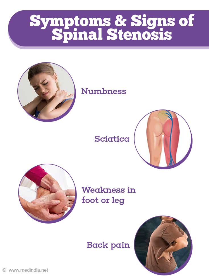
- Numbness or tingling sensation in hand or arm
- Pain radiating from neck down to shoulders, arms and hand
- Weakness in arms and legs
- Problems with walking and balance
- Numbness or tingling sensation in foot or leg
- Weakness in foot or leg
- Pain or cramping in one or both legs on walking or standing, which usually eases on resting, sitting
- Pain in buttock radiating down thighs into calves (sciatica)
- Back pain
- In severe cases, bowel or bladder symptoms (urinary urgency and incontinence)
- X-rays - An X-ray of the spine will show bony changes, such as bone outgrowths that may be narrowing the space within the spinal canal. X-ray test involves a small exposure to radiation.
- Magnetic resonance imaging (MRI) - An MRI scan employs a powerful magnet and radio waves to produce detailed cross-sectional images of the spine. It can pick up damage to intervertebral disks and ligaments, presence of tumors and other lesions. Most important, it can delineate precisely where the nerves in the spinal cord are being pressed
- CT or CT myelogram - In persons who are unable to have an MRI, the doctor may recommend a computerized tomography (CT), scan - a test that combines X-ray images taken from many different angles to give detailed, cross-sectional images of our body. In a CT myelogram, the CT scan is done following injection of a contrast dye. The dye outlines the spinal cord and nerves, herniated disks, tumors and bone spurs
- Doppler ultrasound – A non-invasive procedure that uses sound waves to evaluate blood flow in the limb arteries to rule out peripheral vascular disease as the cause for pain and weakness
- Self-care - Weight management, maintenance of correct posture and erect spine, cessation of smoking, changing standing and sitting habits, correct manner of lifting weights and bending to avoid back strain
- Physiotherapy - Learning and doing exercises to strengthen leg, back and stomach muscles. Spine stretch exercises
- Medication
- Nonsteroidal anti-inflammatory (NSAIDs) drugs - such as aspirin, naproxen, ibuprofen for pain relief and to reduce inflammation, analgesics such as acetaminophen. Longterm use of painkillers may lead to stomach ulcers, liver and kidney problems and are not advised
- Steroids – have excellent anti-inflammatory properties. They may be administered as oral tablets or injections within the affected area of spine for severe pain
- Epidural injections – Injection of steroid and anesthetic (pain numbing) agent within the epidural space of the spinal cord. Nearly half the patients notice some improvement in symptoms following epidural injections although it may be shortlived. These can be repeated upto three times a year if found to help the patient
- Facet injections - Injection of steroid and anesthetic (pain numbing) agent to the painful facet joint to reduce pain and inflammation
- Chiropractic treatments – Application of pressure over the spine to improve bone alignment and to restore normal movement. This treatment may provide relief from pain, reduce muscle spasm and tightness and formation of scar tissue (fibrosis)
- Alternate therapies – Acupressure, acupuncture, biofeedback therapy, nutritional supplements may offer pain relief and improvement of overall health as well
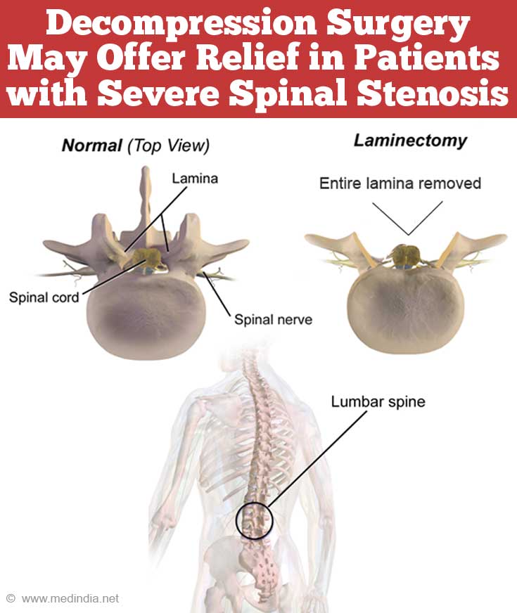
- Spinal decompression (laminectomy) - Under general anesthesia, the arched portion of the vertebra (lamina) is removed and thickened ligaments or bone spurs are removed. Enlarged facet joints may be trimmed and herniated disks may be removed (diskectomy). In patients with severe spinal stenosis, decompression surgery may offer relief in upto 80 percent of cases.
- Spinal fusion surgery – In patients with spinal segment instability, slippage of one vertebra over another, a fusion operation may be helpful. This is often combined along with a laminectomy procedure. Bone grafts (from the hip or a bone bank) may be placed in the area where the lamina was removed. Over 3 to 6 months, the bone grafts will unite the vertebrae into one solid length of bone. Metal plates and screws may be placed to immobilize the area while the fusion occurs.
- Interspinous process spacers – A device is placed between the spinous process of adjacent vertebrae. This device opens up the spinal canal and relieves the pressure. It is a relatively new procedure and longterm outcome is not known. It is a minimally invasive procedure that can be done under local anesthesia.

What Happens in Spinal Stenosis?
Typically, as part of the normal aging and wear and tear, degenerative changes occur in the spine, particularly in the lower back and neck. This may cause partial compression (stenosis) of the vertebral canal within the spinal column. This is referred to as central stenosis.Sometimes there is stenosis of the smaller intervertebral foramina on the sides (refer figure). This is called foraminal stenosis.
What are the Types of Spinal Stenosis?
The types of spinal stenosis are classified according to where along the spine the narrowing occurs. A person can have more than one type. The two main types of spinal stenosis are:
What are the Signs and Symptoms of Spinal Stenosis?
Although there may be evidence of spinal stenosis on CT or MRI scan, many of these patients remain asymptomatic. The nature of symptoms depends on the location of the stenosis and which spinal nerves are affected. These typically begin gradually and progress with time if not treated.
In the neck (cervical spine)
In the lower back (lumbar spine)

How do you Diagnose Spinal Stenosis?
To diagnose spinal stenosis, the doctor will take a detailed history of your symptoms, and conduct a complete physical examination including a neurological examination. He may request several imaging tests such as CT and MRI scans to find a possible cause for the symptoms.
Imaging tests
How do you Treat Spinal Stenosis?
Treatment of spinal stenosis depends on the location of the stenosis and the severity of symptoms. The condition cannot be halted or cured but symptoms may be controlled. Initially conservative measures will be recommended to try and manage mild to moderate spinal stenosis. Severe spinal stenosis may need surgery
Non-Surgical Measures
Surgical Treatment
Surgery may be done for nerve root decompression, removal of bony spurs, stabilization of an unstable spine segment causing stenosis


