- Ectopia cordis - (https://radiopaedia.org/articles/ectopia-cordis)
- Conditions and Symptoms - Ectopia Cordis - (https://www.childrenscolorado.org/conditions-and-advice/conditions-and-symptoms/conditions/ectopia-cordis/)
- Introduction - Ectopia Cordis - (https://www.ncbi.nlm.nih.gov/pmc/articles/PMC3762021/)
- A Rare Case Report of Thoracic Ectopia Cordis: An Obstetrician's Point of View in Multidisciplinary Approach - (https://www.ncbi.nlm.nih.gov/pmc/articles/PMC5120199/)
- [Congenital anomalies of poor prognosis. Genetics Consensus Committee]. - (https://www.ncbi.nlm.nih.gov/pubmed/27234469)
- Ectopia Cordis with Associated Foetal Anomalies: Antenatal Ultrasonographic Detection - (http://www.jkscience.org/archive/Volume14/Ectopia%20Cordis%20With%20Associated.pdf)
- Prenatal sonographic diagnosis of ectopia cordis - (https://www.ncbi.nlm.nih.gov/pubmed/10477886)
- What is "ectopia cordis"? - (http://signssymptoms.org/definition-for-disease-signs-symptoms-and-treatment/?ectopia_cordis-1163)
What is Ectopia Cordis?
Ectopia Cordis [EC] is an extremely rare birth defect in which the heart is abnormally located either partially or totally outside the chest cavity. Normally the heart is located in the chest cavity in between the lungs. But in ectopic cordis the heart forms either partly or totally outside the chest cavity. The ectopic heart may protrude through the neck, chest, or abdomen. In most cases, the heart protrudes outside the chest through a split breast bone [sternum]. This birth disorder is mostly fatal and a new born with this condition rarely survives for more than a few hours or days.
During a baby’s development in the mother’s womb, the chest wall does not fuse together as it normally should. This prevents the heart from developing in normally, leaving it exposed outside the chest wall.
What are the Classifications of Ectopic Cordis?
Ectopia cordis is derived from Latin, ectopic meaning “outside or out of place”, and cordis meaning “heart”. Ectopia cordis is also classified in two different ways according to its location.
With reference to the chest cavity
- In partial ectopia cordis, the heart is located outside the chest wall just under the skin and the heart can be seen beating through the skin.
- In complete ectopia cordis, the visible heart is situated totally outside the chest, with only a thin membrane to covering it.
With reference to the vertebral column
- Cervical ectopia cordis- if the heart is placed in the neck area. This occurs in 3 percent of cases.
- Thoracic ectopia cordis- if the heart is in the thoracic cavity. This is the most common placement and is seen in 53 percent cases.
- Thoracoabdominal ectopia cordis- if the heart is placed between the abdominal and thoracic cavities. This placement is seen in 10 percent of cases.
- Abdominal ectopia cordis- if the heart is placed in the abdominal cavity. This is seen in 30 percent of cases.
Facts on Ectopia Cordis
- Ectopia cordis occurs in 5-8 per one million live births.
- It is more common in male infants.
- Among the various types, abdominal ectopic cordis has a better prognosis while cervical and thoracic ectopia cordis are quite fatal within days.
What are the Causes of Ectopia Cordis?
The cause of ectopia cordis is not known. During fetal development, the breastbone [sternum] does not develop properly. This leads to abnormal placement of the heart in the fetus. The pathology behind ectopic cordis could be due to improper alignment of the mesoderm (one of the three germ layers in a very early embryo) at the midline. Some theories suggest chromosomal abnormalities. Studies indicate that damage or rupture of the amniotic sac in early stages of pregnancy can cause fibrous bands of amnion [the inner membrane of an embryo] leading to deformities of the heart. This is also known as amniotic band syndrome. Other theories suggest an intrinsic defect of the blood circulation called the vascular disruption theory or a fault during fetal folding process.
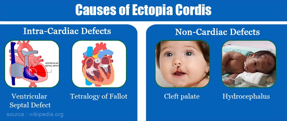
Ectopia cordis is usually associated with other congenital defects.
Intracardiac defects:
- Ventricular septal defect (common) - a hole between the ventricle or lower chambers of heart
- Tetralogy of Fallot (common) -includesventricular septal defect, pulmonary stenosis, (narrowing of the artery that connects the heart with the lungs), an overriding aorta, and right ventricular hypertrophy
- Atrial septal defect - a hole between the atrium or upper chambers of heart
- Tricuspid atresia - absence of the tricuspid valve
- Double outlet right ventricle - where the great arteries connect to the right ventricle
Non-cardiac defects:
- Omphalocele (common) - a rare abdominal wall defect in which the intestines, liver, and occasionally other organs remain outside of the abdomen in a sac.
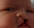
- Cleft palate - a birth defect that occurs when a baby’s lip or mouth does not form properly during pregnancy.
- Skeletal dysplasia - is a complex group of bone and cartilage disorders that affect the fetal skeleton.
- Hydrocephalus -fluid built up in brain.
- Hypoplastic lung disease - is a failure of development of the lungs in a fetus. There is less blood flow and inadequate gas exchange which may lead to electrolyte imbalance in the body [dyselectrolytemia].
- Meningocele - protrusion of the membranes that cover the spine through a bone defect in the vertebral column.
Pentalogy of Cantrell: The thoracoabdominal ectopic cordis type can be present along with other associated anomalies that involve the diaphragm, abdominal wall, pericardium (membrane that lines the heart), heart and lower sternum.
- Thoracoabdominal ectopic cordis with lower sternum cleft
- Omphalocele
- Anterior diaphragmatic hernia
- Diaphragmatic pericardium defects
- Congenital intracardiac defects like ventricular septal defect or Tetralogy of Fallot
What are the Symptoms and Signs of Ectopia Cordis?
This condition is immediately apparent at birth. Along with location of heart outside the body the child may experience difficulty in breathing or appear bluish (cyanotic) due to inadequate oxygenation of the blood.
- Babies with ectopia cordis may also have cleft palate, spinal defects, and improper development of lungs.
- Most of the ectopia cordis patients have excessive curvature in the upper back causing a round back or hunchback.
How do you Diagnose Ectopia Cordis?
Ectopia cordis can be readily diagnosed in utero as early as the first trimester or in the second trimester of pregnancy by about the 10th or 11th week thanks to the advent of ultrasound. If not discovered during pregnancy, it becomes obvious as soon as the baby is born.
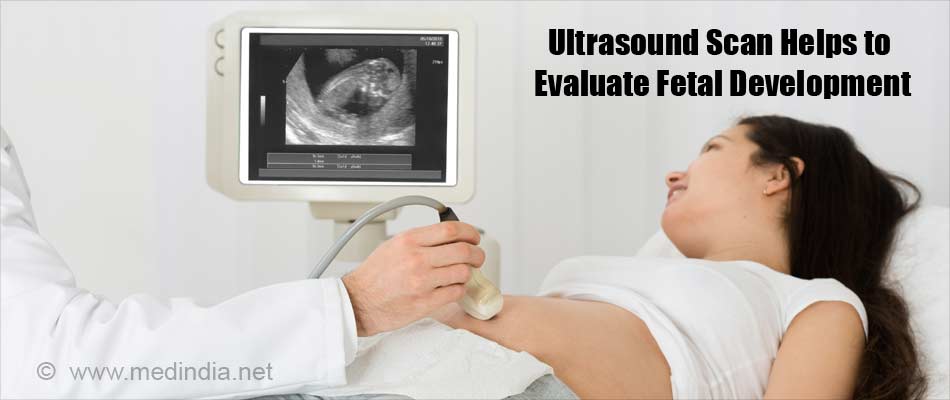
- Ultrasound - imaging uses sound waves to produce pictures of the fetus inside of the body. Ultrasound scans are used to evaluate fetal development. It is safe, noninvasive, and does not use ionizing radiation. Three-dimensional ultrasound along with Doppler gives an accurate early diagnosis.
- Plain radiograph - will confirm the abnormal location of the heart and defect in the breast bone [sternum] in an infant along with other skeletal anomalies.
What are the Complications of Ectopia Cordis?
If the heart is positioned completely outside the body the heart is extremely vulnerable to injury and infection.
The associated medical problems that most infants born with ectopia cordis face such as atrial septal defect, ventricular septal defect, cleft palate, spinal defects, imperfectly formed lungs, and meningocele may cause poor circulation, difficulty in breathing, low blood pressure and electrolyte imbalance.
How do you Treat Ectopia Cordis?
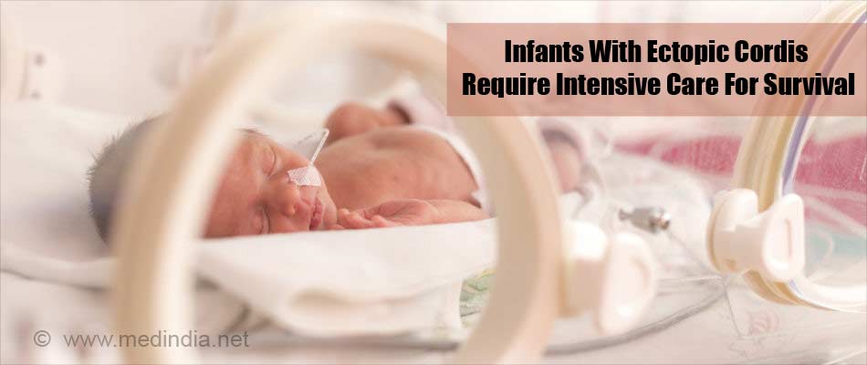
Infants who survive birth with this condition require intensive care. This may include incubation and use of a respirator. Sterile dressings may be used to cover the heart. Other supportive care, such as antibiotics to prevent infection, is also needed.
Surgery - involves repositioning the heart and providing adequate coverage of the chest wall defect. Additional surgeries may be needed to repair other heart or abdominal wall defects.
What is the Prognosis or Outcome for babies with Ectopic Cordis?
The survival rate for infants with this ectopic cordis is about 10%. Those who survive die within the first few days of life or require extensive surgeries and lifelong medical care delivered by a team of specialists.


