Recent research conducted in Germany indicates that bronchiolo–alveolar carcinoma frequently shows a sonographic pattern of ‘pneumonia’.
The acoustic–physical conditions of the thorax produces the most serious limitations for the sonographic diagnosis of pulmonary diseases.Sonography of pleural-based lesions has been established as a method complementary to conventional X-ray examination. The sonographic pattern of bacterial pneumonia is predominantly well known and characterized by an air bronchogram, a marked vascularity on color Doppler sonography, and a high-impedance arterial flow pattern on Doppler spectral analysis.
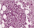
Bronchiolo–alveolar carcinoma (BAC) is defined as a special subtype of adenocarcinoma. Histologically it shows wallpaper like growth within the alveoli, confirmed to the topographical structures of the lung tissue.
The authors set out to describe the sonographic patterns of this tumor entity in seven consecutive patients with peripheral BAC on X-ray examination. Their findings appear in the October issue of the European Journal of Ultrasound. The analysis of data from this study demonstrates a marked vascularity or a vascular tree like pattern in all patients with BAC investigated, which are known to be characteristic for pneumonic or obstructive pulmonary lesions. The arterial spectral analysis of the patients predominantly showed a high-impedance flow in nearly all cases investigated. This flow pattern is also described to be characteristic for pneumonic or obstructive pulmonary lesions and is a well-known flow pattern of pulmonary arteries.
The authors conclude that BAC predominantly shows a pattern of ‘pneumonia’ as demonstrated by conventional B-mode sonography as well as color Doppler sonography.


