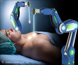
Engineers at the Duke University are working on a technology that may someday enable physicians to use high frequency ultrasound waves both to visualize the heart's interior in three dimensions and then selectively destroy heart tissue with heat to correct arrhythmias.
This is the first time a three in one attempt has been made regarding 3-D imaging, ultrasound and therapy. The researchers believe that the technique may revolutionize the most widely used method for destroying or ‘ablating’ heart tissue that is responsible for irregular heart rhythm.The dual-function imaging-plus-ablation ultrasound probes as small as three millimeters. As many as 200 tiny cables have been fitted into a 3 mm catheter due to the advancement in cable technology. The ablation beam emerging from the 112-transducer array is 50 times more energetic than the imaging beam.
"The nice thing about ultrasound ablation is that you don't have to touch the tissue. The sound waves propagate through the blood and can ablate the tissue from a distance of a centimeter or two", according to the engineers who have developed the technique.
The current available technique for the purpose is based on radio waves emitted from the end an electrode probe. The radio waves destruct the desired tissue upon contact by the production of excessive heat. Moreover the fluoroscopic imaging used to locate the device produces a very poor quality image, often with a fuzzy background.
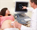
The engineers have previously pioneered techniques rendering the kind of soft tissue internal images that enable fetuses to be seen in the womb. They have also pioneered the use of ultrasound to create 3-D images of the heart and other organs.
During the past five years several tiny internal ultrasound-imaging probes have been developed that offer a significant advantage over X- rays. The acquisition of only two-dimensional images by these probes limits the use of the probe for tissue ablation.
Advertisement


