"Clinically, scientists have focused on trying to understand how cartilage and joints degenerate in osteoarthritis. But no one knows why it hurts," said Dr. Anne-Marie Malfait, associate professor of biochemistry and of internal medicine at Rush, who led the study.
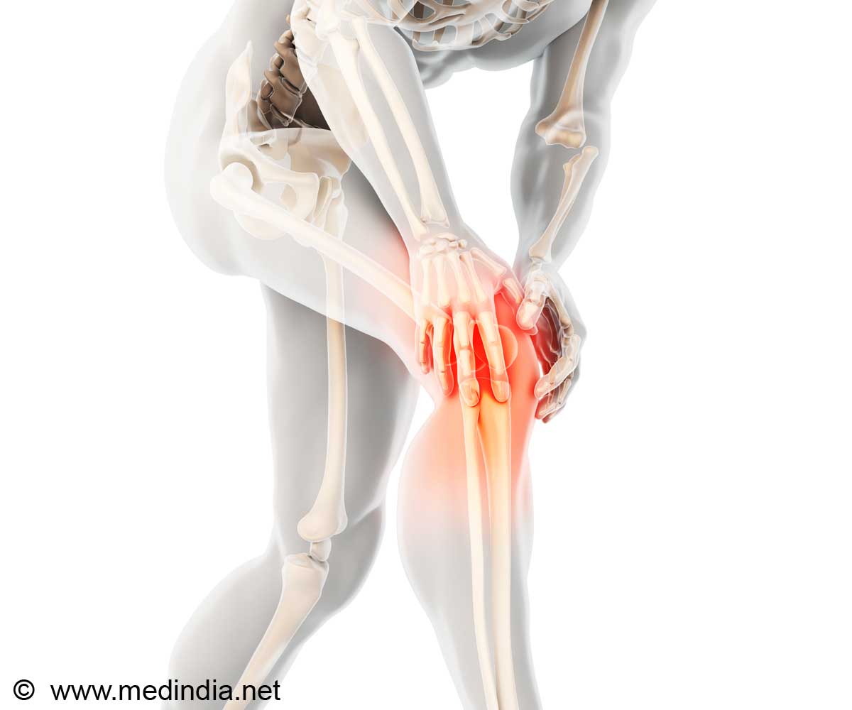
Joint pain associated with OA has unique clinical features that provide insight into the mechanisms that cause it. First, joint pain has a strong mechanical component: It is typically triggered by specific activities (for example, climbing stairs elicits knee pain) and is relieved by rest. As structural joint disease advances, pain may also occur in rest.
Heightened sensitivity to pain, including mechanical allodynia (pain caused by a stimulus that does not normally evoke pain, such as lightly brushing the skin with a cotton swab), and reduced pain-pressure thresholds are features of OA.
Malfait and her colleagues took a novel approach to unraveling molecular pathways of OA pain in a surgical mouse model exhibiting the slow, chronically progressive development of the disease. The study was conducted longitudinally, that is, the researchers were able to monitor development of both pain behaviors and molecular events in the sensory neurons of the knee and correlate the data from repeated observations over an extended period.
"This method essentially provides us with a longitudinal 'read-out' of the development of OA pain and pain-related behaviors, in a mouse model" Malfait said.
Advertisement
Monocyte chemoattractant protein-1 regulates migration and infiltration of monocytes into tissues where they replenish infection-fighting macrophages. Previous research has shown that MCP-1/CCR2 are central in pain development following nerve injury.
Advertisement
This result correlated with the presentation of movement-provoked pain behaviors (for instance, mice with OA travelled less distance, when monitored overnight, and climbed less often on the lid of their cage - suggesting that they avoid movement that triggers pain), which were maintained up to 16 weeks.
Mice that lack Ccr2 (knockout mice) also developed mechanical allodynia, but this began to resolve from eight weeks onward. Despite having severe allodynia and structural knee joint damage equal to that in normal mice, Ccr2-knockout mice did not develop movement-provoked pain behaviors at eight weeks.
To confirm the key role of CCR2 signaling in development of the observed movement-provoked pain behavior after surgery, the researchers administered a CCR2 receptor-blocker to normal mice at nine weeks after surgery and found that this reversed the decrease in distance traveled, that is, movement-provoked pain behavior.
Interestingly, levels of MCP-1 and CCR2 returned to baseline or lower by 16 weeks in mice exhibiting movement-provoked pain behaviors. This finding may suggest that the MCP-1/CCR2 pathway is involved only in the initiation of changes in the DRG, but once macrophages are present, the process is no longer dependent on increased MCP-1/CCR2.
"Increased expression of both MCP-1 and its receptor CCR2 may mediate increased pain signaling through direct excitation of DRG neurons, as well as through attracting macrophages to the DRG," the researchers said.
"This is an important contribution to the field of osteoarthritis research. Rather than looking at the cartilage breakdown pathway in osteoarthritis, Dr. Malfait and her colleagues are looking at the pain pathway, and this can take OA research in to a novel direction that can lead to new pain remedies in the future," said Dr. Joshua Jacobs, professor and chairman of orthopedic surgery at Rush University Medical Center.
A paper describing the research was recently published in the print version of the Proceedings of the National Academy of Sciences.
Source-ANI


