Highlights
- Pancreatic cancer is often detected only in advanced stages with poor survival rates.
- All living cells cells emit extracellular vesicles (EV’s) which are tiny bubbles of material
- Pancreatic cancer cells emit EV’s carrying a surface protein EphA2-EV that is a biomarker for pancreatic cancer.
- Detection of EphA2 levels in blood using nanoparticle probe helps early diagnosis and monitoring of treatment.
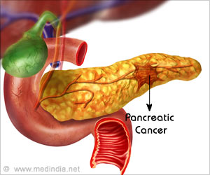
Pancreatic cancer remains difficult to diagnose in its early stages as it is very often asymptomatic. The tumor grows aggressively and is fairly advanced by the time it is diagnosed clinically. As a result, prognosis remain poor and nearly 80% of patients diagnosed, rarely live for more than a year following diagnosis.
This research describes a mechanism by which it is possible to diagnose pancreatic cancer in its early stages by measuring the blood levels of a protein EphA2. This protein is secreted by the tumor and increased levels can be detected in blood and serves as a biomarker for early diagnosis.
Prior to this research, there have been no simple methods to isolate and analyze specific extracellular vesicles making it impossible to use in the diagnostic setting. In addition, the methods used were not able to reliably distinguish tumor derived EV’s from other conditions.
"Pancreatic cancer is one type of cancer we desperately need an early blood biomarker for," Hu says. "Other technology has been used for detection, but it doesn't work very well because of the nature of this cancer. It's really hard to capture an early diagnostic signal when there are no symptoms. It's not like breast cancer, where you may feel pain and you can easily check for an abnormal growth."
What are Extracellular Vesicles?
Earlier it was thought that extracellular vesicles merely represented cell debris, but recent studies have shown that they are important structures with a variety of significant functions.
- Transfer of nucleic acids, lipids and proteins between cells, causing physiological as well as pathological alterations in both the parent as well as the target cells.
- Important role in innate and adaptive immunity
- EV’s play an important role in the formation and spread of several cancers including pancreatic cancer.
- Promote development of resistance to anti-cancer drugs by exporting them away from the tumor site or by neutralizing them.
Detection of EV’s Using Nanoparticle Probes
In the simple and rapid technique developed by the research team, a small sample of blood (around 1 microliter), was diluted and applied on a sensor chip coated with antibodies to an EV membrane protein.
The EV bound to the chip by antibody was then added to a mixture of antibody coated two types of nanoparticles, one a red nanorod and the second a green nanosphere. Both these particles were capable of recognizing a membrane protein EphA2 associated with pancreatic cancer.
Due to the close contact between the 2 types of nanoparticles on the EphA2 protein, it led to a coupling effect and change in color and enhancement of the intensity of refracted light. This is visible as a clear signal when examined using a darkfield microscope.
Results of the Analysis
The method was able to predict blood samples from pancreatic cancer with high degree of sensitivity, including those with early stage disease. Additionally it was able to distinguish them from samples of pancreatitis patients and healthy individuals.
In addition, as this method detected slight alterations in EphA2-EV blood levels in pre- and post-therapy blood samples it can potentially be used as a biomarker to monitor response to therapy.
Scope of the Research
- Development of a potential non-invasive, rapid and reliable method to detect early pancreatic cancer
- Monitoring response to therapy in pancreatic cancer
- This technique can also be used to diagnose a wide range of disease conditions by simply customizing the nanoprobes to EV-specific probes for the condition under investigation.
In conclusion, detection of extracellular vesicles, once discarded as mere cellular waste is now showing a lot of promise in the diagnosis and treatment of several diseases. The role of EV’s in drug delivery or as therapeutic agents is being widely investigated by scientists.
Source-Medindia


